Obstructive sleep apnea has been associated with floppy lid syndrome, glaucoma, non-arteritic anterior ischemic optic neuropathy, retinal vascular occlusion, central serous retinopathy, papilledema, and exacerbation of diabetic retinopathy.
Obstructive sleep apnea (OSA) is a sleep-induced breathing disorder characterized by the dangerous cessation of breathing. This condition is often linked to a range of health issues such as diabetes, obesity, hypertension, and cardiovascular disease. Notably, OSA can exacerbate existing health problems, including those related to eye health.
The eye’s blood circulation relies on a delicate network of tiny blood vessels. The retina, optic nerve, and supportive eye structures are particularly sensitive to changes and fluctuations in blood supply. When breathing stops or decreases during sleep apnea episodes, the blood experiences oxygen deficiency and an increase in carbon dioxide partial pressure. These changes can have adverse effects on eye health, highlighting the significant impact of OSA on ocular well-being.
The cycle of sleep apnea is:
- Sleep relaxes the muscle tone of the upper airway and jaw,
- the soft tissues of the upper airway collapses (partially or completely),
- breathing is halted or partially obstructed for seconds to minutes,
- neurosensory feedback to the brain, awakes the respiratory system to breath,
- the individual awakes partially or completely, accompanied by choking , snoring, snorting , and/or gasping,
- breathing resumes.
- The cycle begins again as the person falls back to sleep and repeats multiple times each hour.
Critical to the eyes is that period of decreased blood supply and oxygen. The continuous, chronic decrease in oxygen (and nutrient) supply results in a number of potentially sight threatening eye diseases.
How sleep apnea affects the eyes.
Sleep apnea is a risk factor for the development of several eye diseases or it can make existing eye conditions worse. It can affect the eye from front cornea and conjunctiva, to the back, retina and optic nerve.
The front of the eye: Floppy lid syndrome.
Floppy lid syndrome is a condition characterized by the upper eyelid’s lack of rigidity or tautness to the eyeball, making it elastic-like and easily manipulated. During sleep, the eyelid may spontaneously evert (flip up, turn ‘inside out’) due to the looseness of the upper eyelid.
The primary function of the eyelids is to protect and keep the eye lubricated. However, when an eyelid turns upward, it can lead to dryness, irritation, redness, and chronic conjunctivitis. It’s important to note that sleep apnea is not the sole cause of floppy lid syndrome, and not everyone with OSA develops this condition. Common risk factors include obesity, hypertension, and diabetes.
The optic nerve: Glaucoma.
Glaucoma encompasses a category of eye diseases often associated with elevated eye pressure. Left untreated, it can lead to vision impairment and eventual blindness.
Obstructive Sleep Apnea (OSA) poses a risk factor for the development of Primary Open Angle Glaucoma (POAG). POAG is typically diagnosed in a doctor’s office when elevated eye pressure is detected.
Additionally, OSA is linked to another form of glaucoma where the doctor may observe normal eye pressure but evidence of vision loss due to glaucoma is present. This insidious variant is known as Normal Tension Glaucoma (NTG).
The progression of normal tension glaucoma is believed to stem from decreased blood circulation, leading to damage to the retina and optic nerve during episodes of apnea (Ref :Review of Optometry ) .
Both primary open angle glaucoma and normal tension glaucoma are chronic and progressive causes of vision loss.
The Optic Nerve: Non-arteritic Anterior Ischemic Optic Neuropathy (NAION)

Non-arteritic anterior ischemic optic neuropathy (NAION) is characterized by a sudden, painless loss of vision. Many individuals wake up from sleep with dim or blurred vision as an initial symptom. This condition results in some degree of visual acuity loss and visual field loss, the extent of which can vary.
The cause of NAION is believed to be the compromised blood circulation around the optic nerve of the eye.
NAION is highly associated with sleep apnea, often accompanied by other risk factors such as diabetes, hypertension, and high cholesterol. These factors together contribute to the development and progression of this condition. (Ref:: ncbi.nnlm.nih.gov website )
)
The optic nerve: Papilledema
Papilledema refers to the swelling of the optic nerve head as it enters the eyeball, typically caused by an increase in intracranial pressure. There are numerous potential causes of papilledema, necessitating a thorough evaluation to pinpoint whether the elevated cranial pressure stems from OSA or another significant issue like hypertension or a brain mass.
It is believed that the decreased oxygen levels during OSA contribute to changes in the blood vessels within the cranium, resulting in increased pressure along the nerves that supply the retina.
The characteristic vision loss associated with papilledema often manifests as visual field defects. With prompt diagnosis and treatment, the central macular area can be spared from permanent damage. (Ref: ncbi.nlm.nih.gov)
The retina: Retinal Vascular Occlusion (a.k.a. retinal vein occlusion)

A retinal vein occlusion occurs due to an increase in intracranial blood pressure, where airway occlusion slows the blood flow from the eye. This backup of blood causes pressure on the vessels, leading to leakage and bleeding into the retina.
Similar to NAION discussed earlier, retinal vein occlusion results in a sudden, painless loss of vision, often experienced upon awakening from sleep. The extent of vision loss varies depending on the blocked vessels, and unfortunately, this loss is usually irreversible.
Research has highlighted a connection between individuals with retinal vein occlusions and those with OSA. These individuals often have other predisposing factors such as diabetes, hypertension, and vascular disease (atherosclerosis). These factors combined contribute to the occurrence and severity of retinal vein occlusion.
Macula of the Retina: Central Serous Retinopathy
Is there such a thing as a stroke in the eye?
The macula, responsible for our sharpest vision, is the central area of the retina. Central serous retinopathy (CSR) is characterized by fluid accumulation under the macula, resulting in blurry and distorted vision (also known as metamorphopsia).
This condition is often observed in individuals, particularly males, under personal stress who also have OSA. The cycle of wakefulness and sleep in OSA contributes to the production of two hormones: epinephrine and norepinephrine. These hormones increase blood vessel permeability, leading to fluid leakage into the macular area.
Fortunately, vision can often return to near-normal once the fluid under the macula resolves. This highlights the impact of stress and sleep disorders on eye health, particularly in individuals with OSA.
How OSA makes existing medical conditions worse: Diabetic retinopathy
Diabetic retinopathy is a serious complication of diabetes, posing a threat to vision. Diabetes, a condition affecting blood vessels, leads to vessel damage due to elevated blood sugar levels, making the vessels more permeable and prone to leakage. When the tiny blood vessels of the retina leak and bleed, it is termed diabetic retinopathy (DR).
In cases where these vessels are damaged, the retina receives less oxygen, prompting the growth of new blood vessels, known as neovascularization. These fragile vessels also tend to leak and bleed, leading to a condition called proliferative diabetic retinopathy (PDR).
OSA serves as a catalyst for increased proliferative diabetic retinopathy. Individuals with PDR, whose retinas are already experiencing oxygen deprivation, are more susceptible to further vessel growth. The cycle of OSA-induced breathing patterns and oxygen deprivation exacerbates diabetic retinopathy, intensifying its impact on vision.
This highlights the intricate relationship between OSA and the progression of diabetic retinopathy.
Treatment of OSA
Obstructive sleep apnea occurs due to a combination of health and lifestyle factors. A sleep study is designed to guide you and the doctor to determine the best course of action to be taken to reduce this sight threatening disease.
Factors recommended to reduce or eliminate OSA:
- Lose weight
- Exercise
- Stop smoking
- Avoid alcohol before sleep
- Don’t sleep on your back, and
- Use a Positive Airway Pressure (PAP) device .
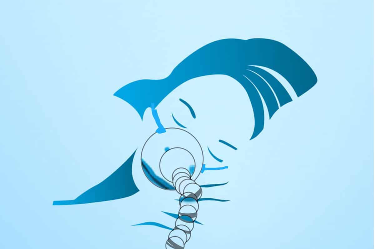
The most commonly prescribed PAP (Positive Airway Pressure) device is the CPAP (Continuous Positive Airway Pressure) device. This device utilizes room air, which passes through a filter and is connected to a humidifier along with a hose and mask. The mask is typically positioned over the nose and mouth.
The CPAP is designed to be used nightly. The gentle pressure of the airflow through the tube and mask works to prevent the collapse of the airway, thus interrupting the cycle of sleep and arousal.
Prescribers often caution patients about potential side effects such as a sore throat, dry nose, and dry eyes. It’s important to note that some individuals may find it challenging to tolerate wearing a mask during sleep.
Eye problems associated with use of a CPAP device
Dry Eyes: The airflow over the eyelids from a CPAP device can exacerbate dryness in susceptible eyes. One solution is to adjust the face mask for a better fit to prevent air leakage. Additionally, using gel-type eye lubricants just before bedtime can help alleviate dryness.
There are goggles specifically designed for use with CPAP devices. The Dry Eye Shop offers moisture retention goggles intended for use with leaky air masks.
Conjunctivitis: Users of CPAP devices may experience redness and irritation in the eyes, with reports of eye irritation and infections. The exact source of bacteria is not entirely clear but is often associated with the use of the mask.
Glaucoma: Research has established a link between Obstructive Sleep Apnea (OSA) and glaucoma. Individuals with glaucoma often experience fluctuations in eye pressure overnight. Studies have indicated that overnight CPAP device use can lead to a higher average eye pressure.
However, there is debate over whether these changes in eye pressure are problematic only for individuals with existing glaucoma or if they pose concerns for those without the condition. This controversy underscores the importance of monitoring eye health while using a CPAP device.
Can CPAP increase eye pressure?
The short answer is that CPAP is generally safe for individuals without glaucoma. However, studies suggest that this may not hold true for those who have already been diagnosed with glaucoma. ( Ref: ncbi.nlm.nih.gov )
In the End…
Sleep apnea doesn’t just impact sleep; it affects overall health. OSA often accompanies other health issues like obesity, diabetes, high blood pressure, and cardiovascular disease. While each of these conditions can affect vision, OSA has been linked to several serious eye conditions that can lead to vision impairment.
It’s crucial for individuals dealing with OSA to recognize that it’s more than just a “bad night’s sleep.” It poses a significant concern for both general health and eye health, emphasizing the importance of proactive management and treatment.

You must be logged in to post a comment.