Photopsia, hallucinations like those in Charles Bonnet Syndrome, double vision, distortions, and halos and starburst patterns around lights are phenomena that individuals with low vision may experience. This comprehensive guide explores these visual disturbances in detail, shedding light on their causes and implications.
- Photopsia
- Hallucinations of Charles Bonnet Syndrome
- Double Vision
- Distortions
- Halos and Starburst Patterns around lights
1. Photopsia
This phenomenon varies from a flash of light to moving patterns of light. These lights appear randomly with no other visual associations. It can occur in one eye or both eyes. They have been seen by those with degenerative eye diseases such as retinitis pigmentosa, age-related macular degeneration, and Stargardt’s disease.
Photopic visual experiences are thought to be residual neurological activity, as they occur in the area of retina which is already damaged. One study associated these photopic experiences with ‘sick retinal cells.’ This would be analogous to the phantom pain experienced by those who have lost a limb. The retinal cell is still firing off without direct light stimulation.
Ref: Photopsia: an often unrecognized symptom …
…
Other types of light flashes not associated with vision loss:
Photopsia experienced by those with vision loss should be differentiated from light flashes associated with retinal detachment or traction on the retina. The cause of the light flash is mechanical stimulation of the retinal cells. These are both active processes that can be a sign of potential vision loss due to retinal detachment or formation of a retinal hole.
Phosphenes are a normal, harmless phenomena of lights seen when the retina of the eye is stimulated. It is also a mechanical stimulation, usually from outside the eye, by rubbing the eye, sneezing, or a punch to the eye, which results in ‘seeing stars.’ The stars are phosphenes.
Ocular migraine aura (aka, retinal, visual, or eye migraines)
Ocular migraines may occur in both those with normal vision and those with low vision.
Signs and symptoms are:
- Area of blocked, ,grey or white vision with scintillating ‘broken shards of glass’ flashing lights,
- starts small in an area and seems to expand until finally dissipating,
- lasts from 5 to 20 minutes,
- usually in one eye, but often can’t tell if it is one or both eyes,
- may happen multiple times in one day.
The causes of ocular migraines are controversial. Most sources indicate that it is thought to be a vasospasm of a retinal blood vessel. The decrease in blood flow to the retina results in the light flashes and areas of transient vision loss. They have no known long term consequences. Do consider a medical evaluation if they reoccur frequently.
2. Vision Loss, Hallucinations, and Charles Bonnet Syndrome
The Charles Bonnet Syndrome (CBS) is that visual hallucination event most commonly assumed to be the phenomenon experienced only by those with low vision.
While the elderly are more likely to experience CBS, there have been reported cases of children with CBS visual hallucinations. The problem with defining CBS is that individuals are not likely to report visual events for fear that they will be perceived as old and ‘losing it,’ or crazy and in need of psychological help or medications.
Those who experience CBS hallucinations are those who:
- Have recent vision loss, not usually those with long term vision loss,
- have lost the vision in both eyes, (20/100 or worse), and
- are mentally alert, attentive, and understand the hallucinations are not real.
Causes of CBS Hallucinations
Studies indicate that those who live alone are more likely to experience the visual images of CBS. The hallucinations may be triggered by sitting quietly or in bed when there is low light levels. It may also be related to fatigue or stress.
The elderly who experience eye diseases of old age most commonly experience CBS, especially those with age-related macular degeneration. CBS can occur for those with vision loss due to cataracts and diseases that affect the retina, such as glaucoma, diabetic retinopathy, optic neuropathy, retinitis pigmentosa or any disease that causes vision loss due to damage along the visual pathway of the brain.
‘Phantom Vision’
It is believed that visual hallucinations of the CBS are the result of the sudden vision loss due to pathology somewhere along the visual system, which includes the eye, the neural connections, or the occipital cortex part of the brain at the back of the head.
The visual hallucinations are analogous to the phantom pain experienced by an amputee. Phantom pain is pain felt in the area where the limb was before it was removed. The brain is filling in the ‘blanks’ where there is no longer any stimulus. The eye doesn’t experience pain with vision loss, but does experiences phantom vision. The nerves of the visual system are still firing, in the absence of stimulus, in the form of images. The Charles Bonnet hallucinations can be called phantom vision!


The hallucinations of Charles Bonnet Syndrome can be simple lines and patterns or complex with animals, people, scenery, and action. Sometimes they are cartoon like, small in size, or grotesque in appearance. They are reported to be brilliant in imagery and blend into the surrounding scenery. They may last a few fleeting seconds or may last for hours. These experiences are episodic and may continue for months to a year. They will eventually end.
Those who have these visual experiences may find them disturbing, but do not usually describe them as horrifying or threatening. The images do not interact with them, they are merely observers.
Getting Rid Of CBS Hallucinations
There are case reports of people, disturbed by the hallucinations, seeking medical help. There is no one direct treatment for these visual disturbances. Resolving the underlying condition of the vision loss will help. For example, cataract surgery for vision loss due to cataracts. Unfortunately, restoring vision is not usually possible.
Reports indicate people have used eye movement, eye closure, or lighting changes at the time to dispel the hallucination. Reducing isolation, vision rehabilitation, and the use of optical aids can help an individual redirect visual stimulation.
3. Distortion of Vision
Often patients with new eye glass prescriptions will complain about images, lines, and print as being curved or not being straight. When they take the glasses off, the distortion disappears. This is a problem often experienced by the normal sighted, who are sensitive to changes in prescription. Those with low vision may experience distortion of images when using low vision magnifying aids. Choosing a different magnifier will solve that problem.
Distortion associated with Eye Disease
Those with low vision can experience distortion due to the underlying disease process that has caused their vision loss. Distortion associated with eye disease may suddenly appear and will persist until the underlying disease process is treated.
Sharp, flat, and distinct images depend on a retina that is intact and flat against the posterior wall of the eye. A retina that is raised or displaced will give to the brain an image that is raised or displaced, in other words, distorted.
Examples of causes of distortion due to disease:
- Fluid under the retina, ex: wet macular degeneration,
- crowding of retinal photoreceptor cells, ex: drusen deposited between the cells,
- areas that appear smaller and crowded, ex: epiretinal membrane.
Changes which cause distortion may be subtle. For those with eye disease, these changes may not be treatable, but are important to monitor to insure they remain stable.
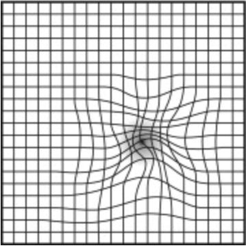
Those with retinal disease are frequently given the Amsler grid to help them monitor for changes in the retina.
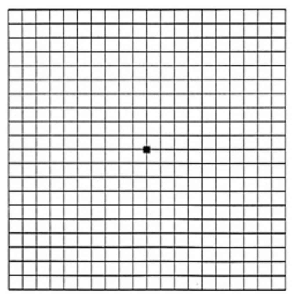
The Amsler grid is simply a piece of paper with a grid of straight horizontal and vertical lines printed on it. With one eye closed, the grid is held 11 inches (28 – 30 cm) from the open eye. Looking straight forward in the direction of the central dot, the patient observes, without looking away from the dot, for any wavy lines or distortion anywhere in the area of their central vision .
To Learn more, see How to Monitor for the Progression of Macular Degeneration
4. Double vision (also known as Diplopia)
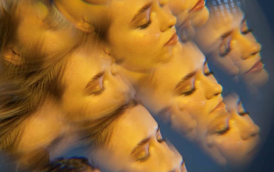
Double vision is seeing two images.
There is double vision which is seen only by one eye called monocular double vision.
Double vision seen when both eyes are open, is binocular double vision.
Normally sighted persons see just one image, because the eyes are perfectly aligned and functioning to maintain a single, clear image. This clear single image helps with depth perception, visual acuity, balance, and movement.
The causes of binocular double vision are varied by the underlying health condition.
- Eye muscle dysfunction. Causes include: inflammation (thyroid eye disease), nerve damage and trauma to eye orbit,
- vascular; causes include stroke, aneurysm, diabetes,
- neurological, causes include: multiple sclerosis, myasthenia gravis, brain lesions,
- decompensated phoria: Misalignments (phorias) of the eyes can be compensated for by the eye muscles of the normally sighted, to maintain a clear single image. Those with vision loss may be unable to maintain eye muscle control for a clear single image resulting in double vision. (Decompensate: unable to compensate for phorias (misalignments of the eyes). ) (Ref: Decompensated Esophoria as a Benign Cause of Acquired Esotropia
 )
)
Astigmatism of the cornea. This is a problem of both normally sighted persons and those with low vision, which is correctable with an eyeglass prescription that corrects for the astigmatism.
Corneal disease will disrupt the smooth refracting surface of the cornea:
- Scarring due to trauma to the cornea,
- Swelling (edema) of the cornea, ex: trauma, infrections,
- Keratoconus is a disease causing progressive thinning of the front surface of the eye. The weaken corneal matrix forms a ‘cone’ which can result in distortion and/or double vision.
Lens of the Eye if disrupted can cause monocular double vision:
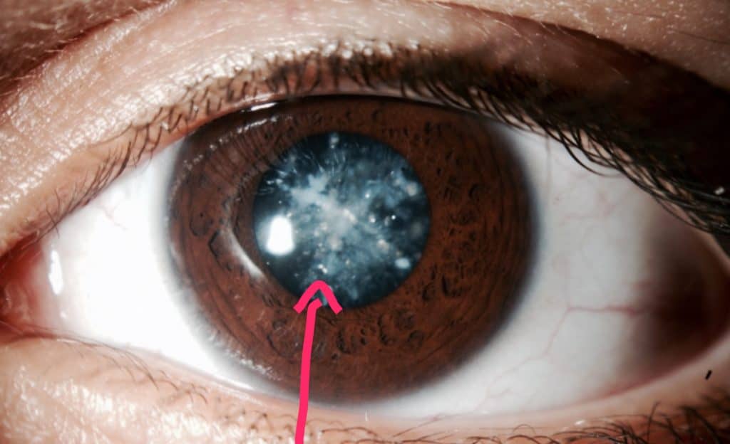
- Cataracts. The lens of the eye should be clear, like a piece of glass. When changes occur to the lens that makes it not clear, these changes are called cataracts. Ex: vacuoles, fluid clefts
Displacement of an intra-ocular lens implant (IOL) in those who have had cataract surgery to remove the natural lens. The plastic implant becomes dislocated, secondary to trauma.
Retinal Disease
Macular swelling (edema) occurs when there is bleeding or fluid leakage into the layers of the retina and macula. This displaces the surface of the retina causing disturbances, primarily blurring and distortion. Doubling or ghosting of images is possible with the accumulation of fluid. Diseases that cause retinal swelling are:
- Wet macular degeneration
- diabetic retinopathy,
- uveitis,
- inflammatory diseases: viral infections, toxoplasmosis,
- auto-immune diseases: sarcoidosis, Behcet’s Syndrome, Vogt-Koyanagi-Harada disease.
5. Halos and Starburst patterns around lights
Causes are:
1. Cataracts are the most common culprit responsible for glare and halos around lights. Other symptoms include blur, dimming of vision, and sensitivity to lights.
The starburst pattern around lights is most notable at night, but may be seen as glare around shiny surfaces during the day. The changes to the matrix of the lens forms surfaces like vacuoles, clefts, splits, and granulations that cause light coming into the eye to be scattered like a starburst or halo.
2. IOLs When a surgeon removes the cataractous lens it is replaced with an iIntraocular lens implant (IOL). Dysphotopsia is a result of light reflecting off the IOL or the edge of the IOL casting a shadow onto the retina. These may appear as glare, starbursts, halos, or darkly blurred areas.
3. Corneal swelling (edema) The structure of the anterior surface of the eye is comprised of different types of cell layers. When the structure of the cornea is disrupted by disease, infection, or trauma, it may start to take on fluids causing swelling. The swollen cornea results in halos, cloudiness, and glare. Examples are:
- Fuch’s corneal dystrophy,
- angle-closure glaucoma attack,
- keratoconus
- occasionally, post-cataract surgery.
In the end…
Although individuals with normal vision may encounter unusual visual experiences, certain disturbances are particularly unique to those with eye diseases. These visual phenomena can serve as warning signs for the onset of a disease process, while others are direct outcomes of the disease itself.
While some treatments can alleviate certain disturbances, it’s important to note that not all visual disturbances are treatable for those with visual impairments. Understanding these effects is crucial for both individuals experiencing low vision and their caregivers, as it helps in navigating the challenges and seeking appropriate care.
