Insight into Low Vision

Recent Posts

Coping with Low Vision: Understanding the Link to Depression and Anxiety


photo of the closed eyes of an ancient statue

Studies have revealed a significant correlation between disabilities, such as vision loss, and the experience of depression and anxiety. Among the various disabilities, vision loss stands out as one of the most daunting, profoundly affecting daily life and playing a pivotal role in the onset of depression and anxiety. The fear accompanying the onset of vision loss often leads to negative attitudes and challenges in adjusting to a life with disability. Understanding this connection is crucial for developing effective coping strategies.

The Impact of Vision Loss on Mental Health: How vision loss can affect your life.

When considering the effects of vision loss, it’s necessary to look beyond the physical challenges and acknowledge the hidden burden it places on mental health. Depression and anxiety frequently accompany vision loss, yet these struggles often go unnoticed and untreated, affecting both adults and children alike.

Depression is a common experience, extending far beyond mere sadness. It can manifest as mild, intermittent episodes or as severe, daily occurrences. Recognizing and addressing persistent, functionally debilitating depression is important for improving overall well-being in those with vision loss.

The American Psychiatric Association inOpens in a new tab.  identifies major depression as having five of the following nine criteria:

1. Depressed mood such as feeling sad, empty, tearful, or irritable most of the day, nearly every day;

2. Decreased interest or pleasure in most activities, most of each day;

3. Significant weight change (5%) or change in appetite;

4. Change in sleep: Insomnia or increased interest in sleep;

5. Change in activity: restless or lethargic;

6. Fatigue or loss of energy;

7. Feelings of worthlessness or excessive or inappropriate guilt;

8. Find it difficult to think or concentrate or an inability to make decisions; and

9. Thoughts of death or suicide, and

I would like to add:

10. Outbursts of anger, irritability, seemingly inappropriate for the situation.

While struggling with vision loss can be emotionally distressing, it’s the ensuing changes in daily functioning that often lead to frustration, depression, and anxiety.

The loss of vision significantly alters one’s quality of life, particularly among older adults. Depression can stem from feelings of diminished self-worth, a loss of independence, and an overall sense of losing control over one’s life.

This decline in quality of life creates a ripple effect on mental health, triggering a cascade of negative conditions. The mental well-being of an individual with vision loss can further diminish quality of life, potentially impacting their overall health and well-being significantly. Understanding these challenges is important for providing holistic care and support for those with visual disabilities.

Prevalence of Depression among the Visually Impaired

As the population of developed countries continues to age, there is a concurrent rise in vision impairments among older adults.

Numerous studies have highlighted a concerning trend: approximately one-third (1/3) of recently impaired adults experience clinical depression. This rate is twice as high as that observed in the same age group without vision impairments. The highest rates of depression are found among those who have recently lost their vision.

Understanding these statistics sheds light on the urgent need for targeted mental health support for individuals facing visual impairments.

Risk factors for depression:

Living alone.  Feeling like a prisoner in your home due to insecurity about moving out of the familiar.  Those with vision loss  often suffer from social isolation.

Financial worries. Those that lose vision may have to stop working. There may be an increase in medical bills. Financial responsibilities may fall on another family member. These changes in the family dynamic can cause additional stress.

Vision loss at a relatively younger age (ages 40 -59)as opposed to older (>60).  Most middle aged people are actively involved in work and activities.  This vision loss is now an unanticipated obstacle and a hindrance to future plans and expectations.

Poor functional vision.  How the individual feels about their vision and how they are able to function affects their mental status and level of depression. The increased level of frustration and depression results in lower visual functioning capability.

Unable to accept vision loss.  Those that live with hope that somehow vision will return, that a treatment exists,  or a miracle cure will arise.  Those who do not accept and move on to adjusting and adapting are susceptible to depression.

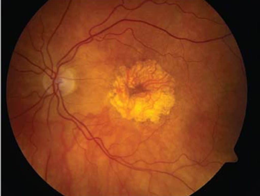
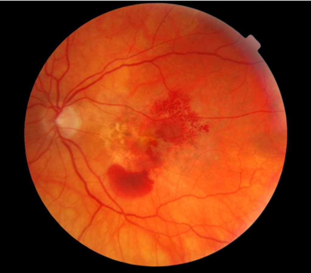
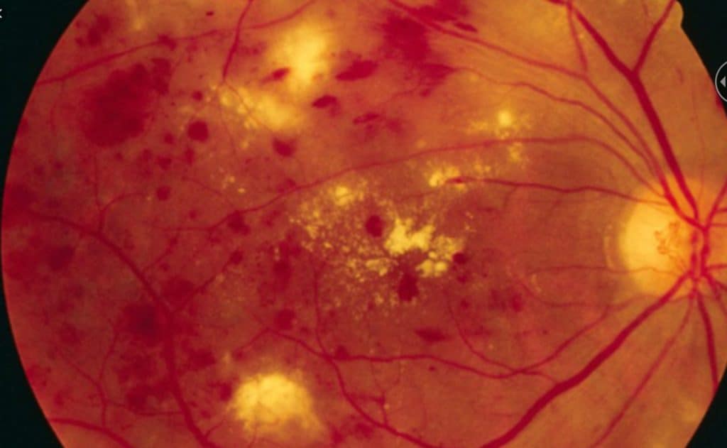
Macular degeneration.  AMD is the most common cause of low vision in adults over the age of 60 in the developed countries. 

Other health problems. Poor physical health in and of itself is a risk factor for depression.  Sight loss can be a compounding factor placing additional restrictions on activates and work.

Ref: Depressive symptoms in people with vision impairment: …BMJ open

Anxiety

Anxiety often walks hand in hand with depression, presenting its own set of challenges. Much like depression, anxiety can persist and manifest in various forms. It encompasses worries, both big and small, affecting behaviors and the willingness to adapt.

Minor fears might deter you from exploring new opportunities. Loss of confidence and frustration over small mishaps, such as pressing the wrong button on a stove or avoiding using a TV remote out of fear of making an irreversible mistake, are common experiences.

On a larger scale, anxiety can lead to isolation and avoidance of social interactions. Fear of social judgment or pity, apprehension about traveling alone, and the dread of social embarrassment due to misunderstood actions can become overwhelming.

The inability to read non-verbal cues, such as body language or facial expressions, adds to this anxiety. Feeling adrift, unaware of subtle details or visual jokes, can create a sense of being “alone in a crowd.” Small embarrassments may amplify the anticipation of others’ judgments regarding one’s capabilities and limitations, leading to avoidance of social situations— a form of self-isolation.

Learn more about low vision and problems with facial recognition in my other article: Why Those with Low Vision have a Problem with Facial Recognition

The greatest fear looms: blindness. Often, others fail to grasp the complexities of this disability, especially when individuals with visual impairments can function normally in most situations (a concept known as ‘passing,’ which I discuss in another article). Falling short of these perceived expectations, even if only apparent to oneself, can significantly impact self-esteem and heighten anxiety levels. Understanding and addressing these anxieties are steps towards holistic well-being for the visually impaired.

In her classic book, Clinical Low Vision, Dr. Eleanor Faye was able to best describe the dilemma of the visually impaired:

“The terms “sighted” and “blind” represent groups possessing well established stereotypes and culturally expected rules of behavior.  The position and role of the partially sighted person is much less clear owing to the tremendous range of variability in partially sighted types.  Generally society views them as sighted and expects them to function as such.”

Want to learn more about ‘passing,’ see my other article: Pretending not to be Visually Impaired: Passing with a Disability

Depression and Anxiety in Visually Impaired Children

Children who acquire low vision may struggle to articulate their concerns or the challenges of their disability to others. Their understanding of their differences may not be clear, adding complexity to their emotional development.

These early years are crucial for self-perception and identity formation. How others treat them plays a pivotal role. The visually impaired child might perceive themselves as ‘slow’ or ‘dim’ due to the way adults, especially those unfamiliar with the disability, may overly assist them. This can lead to a lack of confidence and insecurity about their capabilities.

Sighted children, observing the low vision child’s behaviors such as holding objects close, using magnifiers, or thick glasses, may engage in teasing or ridicule, as children often do. The limitations in participating in activities that sighted children take for granted can further contribute to low self-esteem and self-worth in these young individuals.

Additionally, children are perceptive to their parents’ anxieties. Parents, understandably concerned and seeking assistance, can inadvertently transfer their fear and worry to their child, amplifying the child’s own sense of anxiety and insecurity. Understanding these dynamics is important for providing effective support and fostering positive self-image and emotional well-being in visually impaired children.

Like adults, persistent sadness or persistent fears can be signs of depression and anxiety.  The child may not be able to express their feelings.  Watch for signs of anxiety.

  • Irritability,
  • fear of school and social situations.,
  • fatigue, or
  • trouble sleeping,
  • stomachaches, and
  • nausea.

Signs of depression are similar to adults:

  • Persistent sadness,
  • reluctance to participate in activities,
  • changes in eating patterns – eating a lot more or a lot less than usual,
  • changes in sleep patterns – sleeping a lot more or a lot less than normal,
  • low or overly agitated energy level– being tired and sluggish or tense and restless a lot of the time,
  • having a hard time paying attention,
  • low self-esteem, low self-confidence.

 Ref: Centers for Disease Control and PreventionOpens in a new tab.

Supporting Your Visually Impaired Child Through Depression and Anxiety

Life with a vision impairment is far from the end of the road. Countless stories exist of blind and visually impaired individuals excelling in college, pursuing fulfilling careers, and even competing in the Olympics.

Empower your child to find their own solutions to challenges. Encourage them not to let their disability become an excuse that holds them back. While it’s essential to provide love, affection, and moral support, avoid dominating their life with excessive assistance. Encourage them to connect with groups of friends or support groups to develop social skills and coping mechanisms. With your guidance, they can learn to navigate the challenges they face.

Consider seeking the assistance of qualified therapists who specialize in supporting children with visual impairments. These professionals can help both you and your child gain a deeper understanding of their emotions and provide valuable strategies for managing depression and anxiety. By working together and fostering independence, you can empower your visually impaired child to thrive and embrace a fulfilling life.

 A reader made a recommendation for an article that takes a deeper dive into Teenage DepressionOpens in a new tab.

Addressing Depression in the Visually Disabled: The Treatment Pathway

Treating depression in the visually disabled community can be challenging as it often goes undetected and untreated. There is a reluctance to acknowledge depression, perhaps due to the stigma surrounding mental health issues, or the misconception that it’s an inevitable part of aging with no definitive cure. Some individuals may find it difficult to seek help if they deny their feelings, dismissing them as just temporary sadness.

It takes a village

Successful treatment typically involves a comprehensive approach, starting with a low vision evaluation conducted by a specialized professional such as a low vision specialist, often an optometrist. Additionally, a rehabilitation therapist—usually an occupational therapist trained in low vision rehabilitation—can provide valuable assistance. In some cases, a psychiatrist trained in counseling individuals with disabilities may also play a important role in developing a holistic treatment plan. This multidisciplinary approach ensures that the individual receives the tailored support needed to address their depression effectively.

Tips on coping with vision loss, read my articles: How to Adjust to Vision Loss, 7 Keys to Coping

Low Vision Evaluation

A low vision evaluation serves as a pivotal tool for problem-solving in the journey of those experiencing depression or anxiety due to vision loss. It offers individuals the opportunity to explore and regain functional capabilities, focusing not on treating the disease itself with medications or surgery, but on developing new strategies to adapt to vision loss and enhance the functionality of their remaining vision.

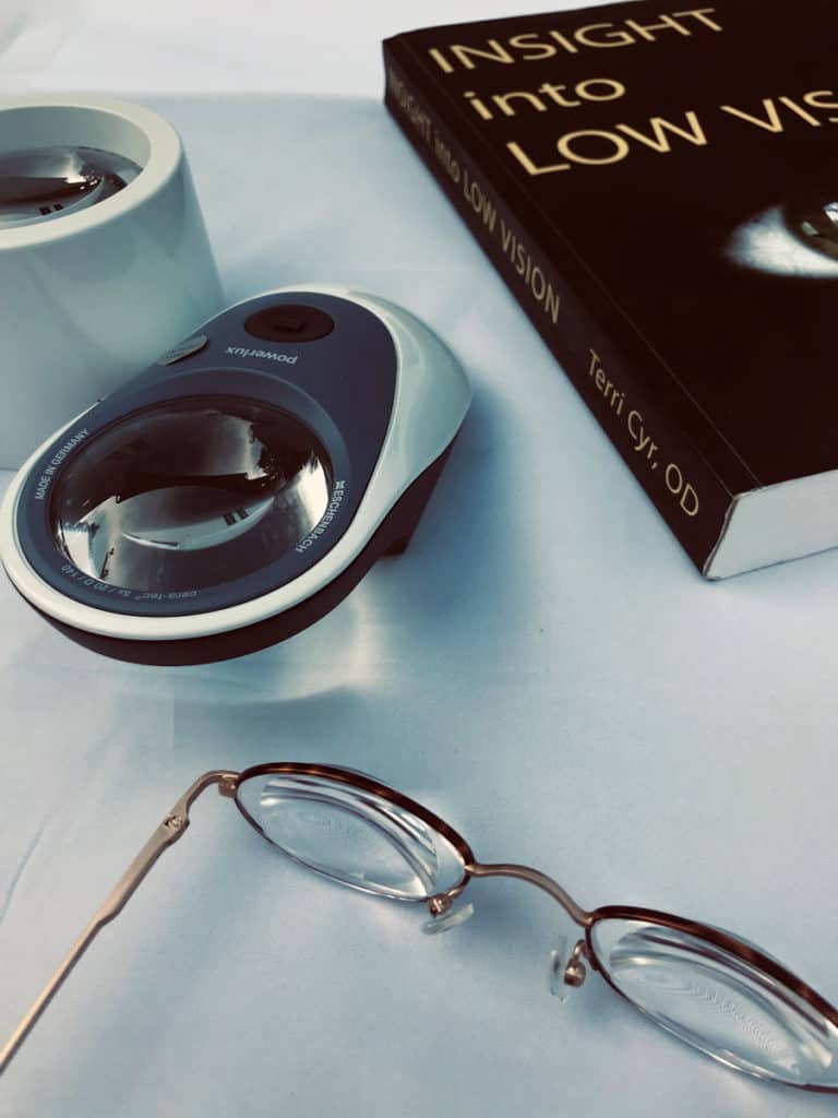
Here are the reasons to get a low vision evaluation;

1. To Advocate for Yourself;

2. Introduction to Visual Aids and Techniques;

3. Education and Counseling, and

4. Appropriate Referrals.

The primary aim of a low vision evaluation is to improve the individual’s quality of life. By increasing functional residual vision, the evaluation helps rebuild self-confidence and motivation for adjustment. This process involves exploring a range of new technologies and both optical and non-optical aids that can empower the visually impaired to regain independence and take more control over their life. Through this evaluation, individuals can discover tools and techniques tailored to their specific needs, facilitating a more fulfilling and independent lifestyle despite their visual challenges.

I like to think that technology makes things easier and makes things possible.

See my other article:

6 Reasons Why the Visually Impaired Refuse Low Vision Aids

As wonderful as all this new assistive technology is, it may not solve all the issues associated with depression and anxiety. 

Rehabilitation: A Path to Renewed Independence

Rehabilitation for individuals with vision loss is a transformative journey aimed at empowering them to navigate life in new ways. This process involves learning innovative skills to promote independence and bolster feelings of self-worth. The goals of rehabilitation must align with the individual’s aspirations, going beyond merely teaching skills to someone who may be struggling with motivation due to depression and anxiety.

The essence of rehabilitation is about more than acquiring techniques; it’s about resetting one’s approach to life. Through this process, individuals discover new ways to accomplish tasks and pursue activities they once enjoyed. By focusing on personalized goals, rehabilitation becomes a catalyst for building confidence and embracing independence.

This holistic approach not only equips individuals with practical skills but also helps them rediscover a sense of purpose and fulfillment in their daily lives.

See my other article : Why the visually impaired resist therapy

Psychotherapy for Emotional Support

Psychotherapy, guided by a qualified therapist, offers valuable support for managing interpersonal challenges and family dynamics. A therapist can assist in identifying and addressing social interactions, helping individuals cope with others’ perceptions of their disability, and navigating feelings of grief and sadness associated with their situation. Therapy serves as an important tool for facilitating the transition to a new life as a person with a disability.

Equally significant is the benefit of family counseling, as the dynamics within a family unit often undergo changes with the introduction of a disability. Living with a disabled family member can place strain on relationships, leading to stress, resentment, anger, or anxiety.

Joint counseling sessions provide a space for both family members and the disabled individual to navigate these changes together. By addressing these emotional challenges collaboratively, families can work towards understanding, acceptance, and improved communication, fostering a healthier and more supportive environment for all involved.

Additional article on fear, depression, anxiety, and secondary gain: Why the Visually Impaired Resist Therapy

In the End;

Depression and anxiety are all too common among individuals with visual impairments, yet they often remain undetected or untreated. Rehabilitation and treatment for vision loss, mental health concerns can easily take a backseat, with the assumption that addressing physical impairments will solve everything. This oversight can leave those struggling with depression and anxiety feeling unheard and isolated.

Advocate for yourself when it comes to mental health services. If these services are not offered to you, seek them out. There is no shame in asking for help. Depression is a recognized aspect of acquired disability, as well as aging, and treatment can be an important step towards regaining a sense of well-being and a fulfilling quality of life.

Remember, your mental health matters just as much as your physical health, and seeking support is a courageous step towards a brighter tomorrow.

photo of a younger hand holding an older persons hand

He is a wise man who does not grieve for the things which he has not, but rejoices for those which he has. Epictetus (Greek Stoic philosopher)

How Sleep Apnea Affects Eyesight


illustration of woman sleeping with CPAP mask

Obstructive sleep apnea has been associated with floppy lid syndrome, glaucoma, non-arteritic anterior ischemic optic neuropathy, retinal vascular occlusion, central serous retinopathy, papilledema, and exacerbation of diabetic retinopathy.
Obstructive sleep apnea (OSA) is a sleep-induced breathing disorder characterized by the dangerous cessation of breathing. This condition is often linked to a range of health issues such as diabetes, obesity, hypertension, and cardiovascular disease. Notably, OSA can exacerbate existing health problems, including those related to eye health.

The eye’s blood circulation relies on a delicate network of tiny blood vessels. The retina, optic nerve, and supportive eye structures are particularly sensitive to changes and fluctuations in blood supply. When breathing stops or decreases during sleep apnea episodes, the blood experiences oxygen deficiency and an increase in carbon dioxide partial pressure. These changes can have adverse effects on eye health, highlighting the significant impact of OSA on ocular well-being.

The cycle of sleep apnea is:

  • Sleep relaxes the muscle tone of the upper airway and jaw,
  • the soft tissues of the  upper airway collapses (partially or completely),
  • breathing is halted or partially obstructed for seconds to minutes,
  • neurosensory feedback to the brain, awakes the  respiratory system to breath,
  • the individual awakes partially or completely, accompanied by choking , snoring, snorting , and/or gasping,
  • breathing resumes.
  • The cycle begins again as the person falls back to sleep and repeats multiple times each hour.

Critical to the eyes is that period of decreased blood supply and oxygen.  The continuous, chronic decrease in oxygen (and nutrient) supply results in a number of potentially sight threatening eye diseases.

How sleep apnea affects the eyes.

Sleep apnea is a risk factor for the development of several eye diseases or it can make existing eye conditions worse.   It can affect the eye from front cornea and conjunctiva, to the back, retina and optic nerve.

The front of the eye: Floppy lid syndrome.

Floppy lid syndrome is a condition characterized by the upper eyelid’s lack of rigidity or tautness to the eyeball, making it elastic-like and easily manipulated. During sleep, the eyelid may spontaneously evert (flip up, turn ‘inside out’) due to the looseness of the upper eyelid.

The primary function of the eyelids is to protect and keep the eye lubricated. However, when an eyelid turns upward, it can lead to dryness, irritation, redness, and chronic conjunctivitis. It’s important to note that sleep apnea is not the sole cause of floppy lid syndrome, and not everyone with OSA develops this condition. Common risk factors include obesity, hypertension, and diabetes.

The optic nerve: Glaucoma.

 Glaucoma encompasses a category of eye diseases often associated with elevated eye pressure. Left untreated, it can lead to vision impairment and eventual blindness.

Obstructive Sleep Apnea (OSA) poses a risk factor for the development of Primary Open Angle Glaucoma (POAG). POAG is typically diagnosed in a doctor’s office when elevated eye pressure is detected.

Additionally, OSA is linked to another form of glaucoma where the doctor may observe normal eye pressure but evidence of vision loss due to glaucoma is present. This insidious variant is known as Normal Tension Glaucoma (NTG).

The progression of normal tension glaucoma is believed to stem from decreased blood circulation, leading to damage to the retina and optic nerve during episodes of apnea (Ref :Review of Optometry ) .

Both primary open angle glaucoma and normal tension glaucoma are chronic and progressive causes of vision loss.

The Optic Nerve: Non-arteritic Anterior Ischemic Optic Neuropathy  (NAION)

fundus photo of an optic nerveenlarged from non arteritic anterior optic neuropathy
non arteritic anterior optic neuropathy

Non-arteritic anterior ischemic optic neuropathy (NAION) is characterized by a sudden, painless loss of vision. Many individuals wake up from sleep with dim or blurred vision as an initial symptom. This condition results in some degree of visual acuity loss and visual field loss, the extent of which can vary.

The cause of NAION is believed to be the compromised blood circulation around the optic nerve of the eye.

NAION is highly associated with sleep apnea, often accompanied by other risk factors such as diabetes, hypertension, and high cholesterol. These factors together contribute to the development and progression of this condition. (Ref:: ncbi.nnlm.nih.gov websiteOpens in a new tab.)

The optic nerve: Papilledema

Papilledema refers to the swelling of the optic nerve head as it enters the eyeball, typically caused by an increase in intracranial pressure. There are numerous potential causes of papilledema, necessitating a thorough evaluation to pinpoint whether the elevated cranial pressure stems from OSA or another significant issue like hypertension or a brain mass.

It is believed that the decreased oxygen levels during OSA contribute to changes in the blood vessels within the cranium, resulting in increased pressure along the nerves that supply the retina.

The characteristic vision loss associated with papilledema often manifests as visual field defects. With prompt diagnosis and treatment, the central macular area can be spared from permanent damage.   (Ref: ncbi.nlm.nih.govOpens in a new tab.)

The retina: Retinal Vascular Occlusion (a.k.a. retinal vein occlusion)

fundus photo showing many areas of hemorrhages and fluid leakage of a central retinal vein occulsion
Central retinal vein occlusion

A retinal vein occlusion occurs due to an increase in intracranial blood pressure, where airway occlusion slows the blood flow from the eye. This backup of blood causes pressure on the vessels, leading to leakage and bleeding into the retina.

Similar to NAION discussed earlier, retinal vein occlusion results in a sudden, painless loss of vision, often experienced upon awakening from sleep. The extent of vision loss varies depending on the blocked vessels, and unfortunately, this loss is usually irreversible.

Research has highlighted a connection between individuals with retinal vein occlusions and those with OSA. These individuals often have other predisposing factors such as diabetes, hypertension, and vascular disease (atherosclerosis). These factors combined contribute to the occurrence and severity of retinal vein occlusion.

Macula of the Retina: Central Serous Retinopathy

Is there such a thing as a stroke in the eye?

The macula, responsible for our sharpest vision, is the central area of the retina. Central serous retinopathy (CSR) is characterized by fluid accumulation under the macula, resulting in blurry and distorted vision (also known as metamorphopsia).

This condition is often observed in individuals, particularly males, under personal stress who also have OSA. The cycle of wakefulness and sleep in OSA contributes to the production of two hormones: epinephrine and norepinephrine. These hormones increase blood vessel permeability, leading to fluid leakage into the macular area.

Fortunately, vision can often return to near-normal once the fluid under the macula resolves. This highlights the impact of stress and sleep disorders on eye health, particularly in individuals with OSA.

How OSA makes existing medical conditions worse: Diabetic retinopathy

Diabetic retinopathy is a serious complication of diabetes, posing a threat to vision. Diabetes, a condition affecting blood vessels, leads to vessel damage due to elevated blood sugar levels, making the vessels more permeable and prone to leakage. When the tiny blood vessels of the retina leak and bleed, it is termed diabetic retinopathy (DR).

In cases where these vessels are damaged, the retina receives less oxygen, prompting the growth of new blood vessels, known as neovascularization. These fragile vessels also tend to leak and bleed, leading to a condition called proliferative diabetic retinopathy (PDR).

OSA serves as a catalyst for increased proliferative diabetic retinopathy. Individuals with PDR, whose retinas are already experiencing oxygen deprivation, are more susceptible to further vessel growth. The cycle of OSA-induced breathing patterns and oxygen deprivation exacerbates diabetic retinopathy, intensifying its impact on vision.

This highlights the intricate relationship between OSA and the progression of diabetic retinopathy.

Treatment of OSA

Obstructive sleep apnea occurs due to a combination of health and lifestyle factors.  A sleep study is designed to guide you and the doctor to determine the best course of action to be taken to reduce this sight threatening disease.

Factors recommended to reduce or eliminate OSA:

  • Lose weight
  • Exercise
  • Stop smoking
  • Avoid alcohol before sleep
  • Don’t sleep on your back, and
  • Use a Positive Airway Pressure (PAP) device .

The most commonly prescribed PAP (Positive Airway Pressure) device is the CPAP (Continuous Positive Airway Pressure) device. This device utilizes room air, which passes through a filter and is connected to a humidifier along with a hose and mask. The mask is typically positioned over the nose and mouth.

The CPAP is designed to be used nightly. The gentle pressure of the airflow through the tube and mask works to prevent the collapse of the airway, thus interrupting the cycle of sleep and arousal.

Prescribers often caution patients about potential side effects such as a sore throat, dry nose, and dry eyes. It’s important to note that some individuals may find it challenging to tolerate wearing a mask during sleep.

Eye problems associated with use of a CPAP device

Dry Eyes: The airflow over the eyelids from a CPAP device can exacerbate dryness in susceptible eyes. One solution is to adjust the face mask for a better fit to prevent air leakage. Additionally, using gel-type eye lubricants just before bedtime can help alleviate dryness.

There are goggles specifically designed for use with CPAP devices. The Dry Eye Shop offers moisture retention goggles intended for use with leaky air masks.

Conjunctivitis: Users of CPAP devices may experience redness and irritation in the eyes, with reports of eye irritation and infections. The exact source of bacteria is not entirely clear but is often associated with the use of the mask.

Glaucoma: Research has established a link between Obstructive Sleep Apnea (OSA) and glaucoma. Individuals with glaucoma often experience fluctuations in eye pressure overnight. Studies have indicated that overnight CPAP device use can lead to a higher average eye pressure.

However, there is debate over whether these changes in eye pressure are problematic only for individuals with existing glaucoma or if they pose concerns for those without the condition. This controversy underscores the importance of monitoring eye health while using a CPAP device.

Can CPAP increase eye pressure?

The short answer is that CPAP is generally safe for individuals without glaucoma. However, studies suggest that this may not hold true for those who have already been diagnosed with glaucoma. ( Ref:  ncbi.nlm.nih.govOpens in a new tab. )

In the End…

Sleep apnea doesn’t just impact sleep; it affects overall health. OSA often accompanies other health issues like obesity, diabetes, high blood pressure, and cardiovascular disease. While each of these conditions can affect vision, OSA has been linked to several serious eye conditions that can lead to vision impairment.

It’s crucial for individuals dealing with OSA to recognize that it’s more than just a “bad night’s sleep.” It poses a significant concern for both general health and eye health, emphasizing the importance of proactive management and treatment.

The 7 Truths about AMD


black and white close up image of an eye

Your risk for AMD increases with age, but it is not inevitable or blinding. It is not curable, but can be prevented. Life is not over with a diagnosis of AMD, there are some treatments available.

Truth #1  The risk for developing macular degeneration increases with age.

Population studies have consistently demonstrated a correlation between visual acuity and age, revealing a higher prevalence of vision impairment and legal blindness as individuals grow older. This trend is most pronounced among those aged 65 and above, with a notable escalation in incidence beyond the age of 75.

According to data from the Centers for Disease Control and Prevention (CDC), an estimated 2.95 million Americans aged 40 and above are affected by age-related macular degeneration (AMD), a figure that rises to 67 million across the European Union. AMD stands as the primary cause of permanent vision impairments among individuals over 65, with a particularly elevated occurrence among those of European descent.

The World Health Organization (WHO) approximates that the United States witnesses 200,000 new cases of AMD annually, while Europe experiences 400,000 new cases per year. Projections from the WHO indicate a global population of 196 million individuals with AMD in 2020, with an anticipated increase of 100 million cases by 2040, bringing the total to 288 million cases worldwide.

Truth #2  Macular degeneration is not inevitable as we age.

Truth #3  It is not inevitable that those with macular degeneration will go blind.

Macular degeneration, while often associated with severe vision impairment, does not invariably lead to complete blindness. However, the classification of blindness can vary depending on the criteria applied.

Is macular degeneration considered legally blind?

The American Medical Association initially outlined a definition of blindness, which was subsequently adopted by entities such as the Social Security Administration for eligibility determinations and by the legal system as a standard definition. Many individuals affected by AMD receive state and government assistance due to their legal designation as blind.

According to the criteria established by these agencies, legal blindness is defined as having visual acuity no better than 20/200 (6/60) in the better eye, even with the best prescription for eyeglasses or contact lenses. It’s important to note that being legally blind does not imply total vision loss or the absence of functional vision.

Consider the nature of age-related macular degeneration: It entails the progressive degeneration of the central area of vision known as the macula, leading to diminished visual acuity.

Our ability to perceive visual acuity relies on the densely packed sensory neurons in the central vision area. This region, characterized by a high concentration of neurons, is particularly susceptible to fluctuations in oxygen and nutrient levels. Consequently, it is the central area of acute vision that is most profoundly affected by various health and environmental factors.

The Stages of Macular Degeneration.

There are stages to ‘dry’ macular degeneration.  It is only the final advanced stages that results in “legal blindness.’  90% of those diagnosed with macular degeneration have the dry form.  10% go on to the vision devastating ‘wet’ form of macular degeneration.

Early ‘Dry macular degeneration:

photo of the retina of a healthy eye
Normal retina: central macula and optic nerve
  • Developed slowly,
  • characterized by white small deposits called drusen,
  • causes some visual acuity loss: 20/20  to 20/40 (6/6 to6/12).

Intermediate ‘dry’ macular degeneration:

fundus photo of drusen of macualr degeneration
Drusen in the macular area, intermediate AMD
  • Slow, progression continues,
  • accumulation of more and larger  drusen,
  • more visual acuity loss; 20/40  to 20/100 ( 6/12 to 6/30).

,

fundus photo of macula with severe central damage
Geographic atrophy , cells of the macula have died, resulting in central vision loss

Atrophic ‘dry’ macular degeneration;

  • End stage of ‘dry’ macular degeneration,
  • death of the retinal cells of the macula,
  • visual acuity, less than 20/200 (6/60), ‘legally blind.’

photo of wet macular degeneration
‘Wet’ macular degeneration characterized by leakage and bleeding

‘Wet’ macular degeneration;

  • A sudden, rapid loss of central vision,
  • inflammatory process, characterized by leakage and bleeding of blood vessels beneath the macula, resulting in scarring.
  • Most severe  devastating loss of vision, visual acuity, less than 20/200 (6/60).

Truth# 4 There is no cure for macular degeneration.

Best supplement for Eyes? Read; What is the Best Supplement to Preserve Eye And Brain Health?

Probably the most commercially successful study was the Age Related Eye Disease Study (AREDS).  This study demonstrated that daily supplementation with a formula of antioxidants,  slowed the progression of intermediate to advanced AND by about 25%.  

Description of the ARED study: How do the AREDS and AREDS 2 Differ?

Can you stop the progression or reverse macular degeneration?

The best you can hope for is to halt or slow the progression of ‘dry’ AMD.  The best weapon available is diet and supplementation with anti-oxidants.

The first thing to do is to adjust your diet to include a variety of healthful, whole foods namely fruits and vegetables. These should be prominent in your diet.  To ensure adequate amounts of anti-oxidants, the minimum supplements to take are:

Vitamin C  Studies have shown that Vitamin C is found in high levels in the eye, and has an effect of preventing free radical formation from UV light.  Take 2000 – 3000 mg / day.

Vitamin E  Also protects against free radical damage from UV light exposure and maintains integrity of the lipid membranes of cells.  Dosage; 400 IU /day

Carotenoids, especially lutein and zeaxanthin. The macula has the highest concentration of these two carotenoids in the body, They have a vital function supporting cell function and scavenging ROS.  Dosage:   lutein 10 mg /day  zeaxanthin 2 mg /day.

This is the minimum.  A diet and supplementation regiment high in vitamins and minerals is necessary to maintain good physical and eye health. You might be interested in this other article: ‘3 Most Commonly Recommended Eye Supplements.’

Treatment of ‘Wet’ AMD

10% of those with ‘dry AMD, go on to ‘wet’ AMD.  Although only 1o% develop the ”wet ‘form, it is responsible for 85% of those who are legally blind from AMD.

  This disease is most commonly found in those with vascular disease. 

 The complicating factor is the growth of abnormal blood vessels underneath the macula.  This growth of new, abnormal blood vessels is called neovascularization.  These blood vessels grow in response to low oxygen levels in the retina.  The fragile blood vessels are susceptible to breakage and leakage, which results in more fluid and blood in the macula.  These pockets of fluid and/or blood are damaging and toxic to the vital cells that support the macula. 

Treatments

The treatment for the’ wet’ form of ARMD requires timely detection and intervention by the eye doctor.  Timely detection of macular changes is usually done by self monitoring. Here is an article on “Monitoring for the Progression of AMD.‘ Treatment during the early stages, although it will not reverse damage already done to the central vision area, can reduce the severity of vision loss.  There are three techniques to halt the growth of abnormal blood vessels, thereby limiting potential damage:

1. Injection Only.  Injection into the eye of a drug that stops the development of new, weak and fragile blood vessels.  The drug is called anti-VEGF.  VEGF (vascular endothelial growth factor) is produced by the oxygen starved tissues of the eye to promote the abnormal blood vessel growth.  Although growing new blood vessels seems like a good idea, these new blood vessels are problematic in that they are fragile.  The anti-VEGF drug injection stops the growth to alleviate the problem. (Drug names: Avastin, Lucentis, Eylea, Beovu)

The injected drug is effective for 1 to 2 months, therefore repeat injections are needed every 4 to 8 weeks. Thus far, anti-VEGF injections have been successful in slowing AMD progression and in some cases, there has even been improvements in vision.

2. Injection and Laser.  The drug (Visudyne®) is injected into the arm and within minutes is taken up by the fragile new vessels of the macula.  The injection is followed by a laser treatment to the affected area of the eye to activate the drug.  The drug then closes off the abnormal blood vessels to stop the growth and subsequent leaking and bleeding. This is called photodynamic therapy.  This treatment is not permanent, and may need to be repeated in three months.

3. Laser Only.  Laser treatment is done around the area of the macula that in effect, destroys the surrounding leaky blood vessels.  It is not likely to be done in the macular area, as it can destroy the sensitive retinal cells above the targeted blood vessels, leaving a little blind spot.  This is called photocoagulation laser therapy.

All three of these treatments are done in the retina specialist’s office on an outpatient basis.

Before you get to this stage, learn how to prevent AMD.

Truth #6  MD is preventable.

Age related Macular degeneration occurs after a lifetime of use and possibly abuse.  The truth for those that have been diagnosed with AMD is that it does not have to progress.

1. Healthy body, healthy eyes.

Macular degeneration often arises from a body that has other health issues.  Hypertension, nutrient deficiency (poor diet and poor digestion), cardiovascular disease, and diabetes have all been implicated as risk factors.  Stricter attention to diet  and monitoring and care of other health concerns can reduce the risk of development and progression of eye disease.

High blood pressure medications can have the effect of depleting water soluble vitamins and minerals.  So consider supplementation with high blood pressure medications.

2. Healthful diet and supplement the rest

Eat a diet high in fruits and vegetables.  Try to incorporate fatty fish (for the fish oils). If you can’t eat fish, consider supplementing with the omega 3’s (DHA and EPA)or flaxseed oil. 

Avoid vegetable oils that are processed, like basic vegetable oil, and corn oil (Olive oil and avocado oil are cold pressed and unrefined. Those are OK.)  Also, avoid processed foods, many contain mono- and polyunsaturated oils.  Avoid high sugar and white flour consumption, both of which are low in nutrients  and high in calories.

Think heart healthy diet; Good not only for the heart, but good for the eyes as well.

3. Stop Smoking.

The chemicals introduced to the body through smoking are toxic to the blood vessels and retina.  Studies have shown that smoking can double the risk for AMD. (click for article: JAMA  Ophthalmology)

4. Wear UV protective sun wear   

 A lifetime of chronic UV and blue visible light exposure is thought to be one of the causes of age-related macular degeneration.   The normal eye, when subjected to intense or chronic UV radiation exposure can suffer photo-chemical changes to its tissues.  A diseased eye may be even more susceptible to UV and ‘near’ UV’ damage. UVA and UVB pass through the atmosphere, even through a cloud cover, and can have a negative effect on our skin and eyes.

5. Get Regular exercise. 

The health benefits of regular exercise to prevent weight gain and degenerative disease is well publicized.  Exercise lowers the risk for cardiovascular disease and improves cognitive function.  Both of these benefits are relative to eye heath.

The increased blood flow to the eyes during exercise and after, provides oxygen and nutrients to the retina and supporting structures.

The American Heart Association recommends;

  • Get at least 150 minutes per week of moderate-intensity aerobic activity or 75 minutes per week of vigorous aerobic activity, or a combination of both, preferably spread throughout the week.
  • Add moderate- to high-intensity muscle-strengthening activity (such as resistance or weights) on at least 2 days per week.

See their full recommendations here: heart.orgOpens in a new tab.

6. Know your risk factors.

The most significant of these are:

  • age
  • positive family history
  • cigarette smoking
  • hypertension

Additional factors include:

  • high intake of refined vegetable oils,
  • high intake of fats,
  • high intake of mono- and polyunsaturated fatty acids,
  • high intake of white flour based food products, and
  • blue irises.

Clearly, some of these factors can be controlled, and others cannot.  It still comes down to healthy diet, healthy body, better vision.

7.  Get regular eye examinations.

Regular eye examination are a proactive approach to eye health.

Whether you have any of the risk factors listed above or not, what we all have in common is that we are getting older.  Often patients don’t take action until they have noticed a change in vision.

Modern examination techniques have been developed to detect retinal and macular changes before patients are aware of a loss of visual acuity. 

It is inevitable that as we reach the ages over 45, we will most likely  need reading glasses.  While this need for eyeglasses, called presbyopia, is not considered a disease, it does indicate that our visual system is aging.  All adults should have a routine eye examinations  every 18 months to 2 years.  Those with special health concerns like diabetes, hypertension, and cardiovascular disease, should be examined every year.  Once changes are identified, which indicate a disease process, examination will be more frequent.

Children should also receive eye examinations.  There are those hereditary macular  dystrophies, such as Stargardt’s Disease, Best’s disease, and juvenile retinoschisis,  that affect children in the first and second decades of life.

Truth #7  Life does not end with the diagnosis of macular degeneration.

Acceptance

Life changes for sure.  The changes are social, work related, financial, and psychological. The heaviest burden of vision loss is psychological. It affects your self-perception and social interactions. Acceptance is the psychological hurdle of understanding that things will never be the same. It is the attitude and understanding of how life will change that will determine success of rehabilitation

Adjustment

Things must be done differently.  When you have a  vision impairment, you start noticing how inconvenient life can be.  Things seem to take longer, require more effort, your responses are slower, and actions need to be more deliberate. 

 Goals for schooling or employment will change.  Activities you once enjoyed may change or be restricted.  The independence you once commanded may now be curtailed.  Adjustment is moving forward over all the hurdles of life.  You may not be accepting of every change or challenge, but you just do the best you can do.

You might be interested in this other article ‘Adjusting to Vision Loss.’

Adaption

Adaption is developing skills and learning to use the tools and technology to get you to keep your life on track.  These are skills, techniques, some optical and others non-optical that will be compensatory.  It’s not easy. You must be willing to learn.  This is by far the best time in history for those with low vision.

The rehabilitation process begins with identifying problems that you may be experiencing, then a program of problem solving is determined:

1. Management of the disease process

Optometrists, ophthalmologists, and primary care physicians  have the job of educating you about the disease and its process.  They help identify risk factors, associated health concerns, and implement treatments plans and monitor for progression.  Get regular eye examinations and maintain a  care regiment with your doctor.

The changes advised by the doctors may be related to lifestyle changes. Remember, diet and exercise; are the key to healthy body, healthy mind, and healthy vision.

2. Counseling

Therapists evaluate  psychosocial issues of the persons psychological well-being and family related concerns. They are then able to provide counseling and referrals for guidance and education.  They may also refer the individual to state and government resources and financial assistance.

3.  Non-optical devices

Initially, those with vision loss find in the simplest things more difficult to do.  These are the activities of daily living (often referred to as ADL.)  Things like cooking, personal grooming, shopping, cleaning, and taking medications.  These tasks can be made easier by implementing basic changes like lighting, increasing contrast and color, talking devices, and identifying by touch.  These are non-optical aids.  Occupational therapists help with setting up your home and work spaces for ease of use and training you in the use of both non-optical and optical aids.  

4. Optical aids

First starts with an optimal pair of eye glasses as prescribed to you by your optometrist or low vision specialist.  Those who  work, go to school, or have specialized interests that require them to be able to see detail, will benefit from magnification devices.  These are either standard magnifying lenses and telescopes or video magnifiers.

A low vision specialist will be able to help guide you through the many different types to help you select one most appropriate for the task.  A rehabilitation therapist (occupational)  will help train you to use these optical devices effectively.

4. Cutting edge technology.

Many with vision impairments have discovered that mainstream technology can be adapted to their needs.  Smart phones, tablets, and virtual assistants like Alexa, Siri, Cortana,and Google Assistant are helpful. 

Smartphone developers incorporated accessibility  functions for large print, audio feedback, and lighting controls.  Tablets have the same features and can be used for large print book reading. Software apps have been designed for navigating, reading aloud, magnification among other things.

Virtual assistants are voice controlled bypassing the need to see a screen or keypad.  You can ask questions (time? date?), search the internet, and make phone calls.  When connected to a smart home, functions of the home can be controlled with out seeing switches and dials.  It can also provide security and notifications by audio alerts

Conclusion

Some aspects of the progression of macular degeneration can be controlled, others cannot.  Arming yourself with the knowledge and a willingness to learn can determine your success to adjusting and adapting. 

Learn about monitoring your macular degeneration in my other article: How to Monitor for the Progression of Macular Degeneration

The pessimist complains about the wind. The optimist expects it to change. The leader adjusts the sails. John Maxwell

Why the Visually Impaired Resist Therapy


image of eyes painted on an outdoor as a mural

When it comes to therapy for the visually impaired, factors such as fear, anxiety, depression, and the concept of secondary gain often play significant roles in resistance.

Rehabilitation therapy empowers those with visual impairments to utilize residual vision alongside non-optical devices, optical aids, and cutting-edge technology for a more fulfilling life. This educational process equips individuals with compensatory techniques and tools, enabling them to pursue their educational, work-related, and daily living goals effectively.

This collaborative effort involves eye care providers, therapists, and the visually impaired individual. However, successful rehabilitation hinges on the willingness of the low vision student to adapt, a process complicated by psychological and social factors.

How Depression Affects the Rehabilitation Process

In the realm of adapting to visual impairment, I believe depression stands as the primary obstacle. Research has consistently shown a strong link between vision loss and depression, particularly among older adults. As highlighted in a JAMA Ophthalmology article, “vision loss is among the most common chronic conditions associated with depression in old age.” ( Ref. article JAMA OphthalmologyOpens in a new tab.

The experience of losing vision often leads to a cascade of frustrations, fears, and anxiety, leaving individuals feeling helpless and mired in low self-esteem. The outlook on life dims, and the weight of a visually impaired existence can plunge one into despair and depression.

My exploration into the acceptance of vision loss has underscored that depression is a natural part of the journey from initial shock and fear to eventual acceptance, adjustment, and adaptation. Detailed insights on this process can be found in articles such as “The Adjustment Process.

However, navigating this journey is far from easy. For some, emerging from the emotional depths of depression may never come. The complexity goes beyond vision loss alone, intertwining with age, other health conditions, relationships, living arrangements, financial stability, and even substance abuse.

Identifying depression is the crucial first step towards transitioning to acceptance. Successful rehabilitation services must address these psychosocial needs.

Referring individuals for counseling to manage depression should go hand-in-hand with learning new adaptive skills for life with a visual disability.

Without this counseling, feelings of helplessness, loss of independence, and low self-worth can significantly impede the rehabilitation process.

How Fear can Make the Visually Impaired Resistant to Therapy

Fear, both big and small, can create significant barriers for the visually impaired in therapy. Big fears encompass concerns about navigating the city safely, maneuvering through complex office buildings or airports, and grasping information without the ability to see details — the worry of “do I look foolish?” On the other hand, small fears creep in with thoughts like “do others perceive me as different?” and “did I miss something? Will I need to explain myself to strangers?”

For many, the intricacies of low vision technology can be daunting, especially if the multitude of buttons and features are unidentifiable or their purpose unclear. This fear often stems from the fear of failure. Similarly, adapting in the kitchen or workplace can seem overwhelming, leading to fears of not being able to learn or making a costly mistake.

Why Anxiety Causes Avoidance

Anxiety often thrives in social settings, particularly among acquaintances or strangers. While interactions with friends and family might not trigger much anxiety, the fear of how others will perceive someone with low vision looms large. Questions arise: Do they truly understand the challenges of low vision? Will I need to repeatedly explain my condition, drawing unwanted attention? The tiresome cycle of explaining legal blindness yet functional sight repeats, bringing anxiety about whether I’ll be seen as I was before losing vision, or if I’ll be pitied, treated differently, or judged as less capable. Should I just try to pretend everything is fine?

This anxiety can lead to avoidance as a coping mechanism. The thought process becomes, “If I avoid certain situations, I’ll feel more comfortable and won’t risk appearing foolish. Since I can’t see, I simply won’t participate.”

The Importance of Goals

Setting goals in the rehabilitation process is a collaborative endeavor. While the therapist provides options and solutions, it’s important for the visually impaired individual to voice their needs for adaptation and express which tasks hold their interest. For instance, someone undergoing training in cooking and laundry may lack enthusiasm if their personal aspiration is to return to gardening and enjoying television.

This collaborative approach ensures that the rehabilitation efforts align with the individual’s passions and aspirations, enhancing motivation and engagement in the process.

image of glasses book, and magnifiers
Tools to adapt to reading

The eye care specialist and therapist  set goals for the rehabilitative process.   Goals are things like:

  • reading
  • writing,
  • cooking,
  • household tasks
  • personal care
  • mobility
  • and the use of technology.

Setting goals in the rehabilitation process is a collaborative endeavor. While the therapist provides options and solutions, it’s crucial for the visually impaired individual to voice their needs for adaptation and express which tasks hold their interest. For instance, someone undergoing training in cooking and laundry may lack enthusiasm if their personal aspiration is to return to gardening and enjoying television.

This collaborative approach ensures that the rehabilitation efforts align with the individual’s passions and aspirations, enhancing motivation and engagement in the process.

The Phenomenon of Secondary Gain

Secondary gain is a psycho-social concept that delves into the perceived benefits of a disability for the individual affected. While a disability undoubtedly presents challenges, secondary gain refers to the subtle sense of satisfaction and advantages experienced by the disabled person due to their condition. It’s where the physical impact of vision loss meets its psychological counterpart.

These benefits can range from small conveniences, such as others taking on tasks or reduced work responsibilities, to significant advantages like financial compensations.

Consider this example illustrating secondary gain for someone with visual impairment: Imagine an individual experiencing central vision loss, struggling to see details like mail, bill payments, and kitchen appliance dials. They share these difficulties with family and friends, who eagerly offer assistance. Despite still having usable vision, this individual begins to appreciate the extra attention and care from loved ones. Over time, they might even unintentionally exaggerate the extent of their disability, inadvertently entrapping their willing helpers as caretakers.

However, intervention can provide new avenues. When their optometrist connects them with an occupational therapist specializing in visual aids and adaptive techniques, newfound independence becomes possible. Suddenly, alternative options for managing these tasks emerge. Yet, this newfound independence might challenge the status quo. Family and friends, realizing the visually impaired person’s potential for regaining self-sufficiency, may withdraw the extra help and attention they were offering.

Secondary gain can take many forms:

  • The disability can be used as a means for work avoidance.
  • It is also a good way to gain sympathy. 
  • It can be used as a means to justify not doing activities they do not want to engage in like cooking, taking the garbage out, watching the children, volunteer work, etc.
  • The disability can be used as an excuse for avoiding social gatherings and activities resulting in a form of self-isolation.
  • The disabled may feel they have a good reason to give away responsibilities and make themselves more dependent.  
  • They may also use their disability to increase interpersonal attachments and receive more care-taking.
  • or use their  disability to control, coerce, or just to get their way.

It’s important to recognize that not all visually impaired individuals utilize secondary gains, consciously or intentionally.

Many low vision patients may not even realize they are benefitting from secondary gains. This isn’t to say that their actions are deliberate or malicious; often, it’s someone close to them or a perceptive therapist who recognizes this pattern. Identifying this phenomenon is crucial, as it sheds light on why an individual might seem to stall in the rehabilitation process.

Keep in mind, while some of these attributes of secondary gain described above can be attributed to behaviors of the visually disability, there could also be underlying medical conditions or mental health disorders at play.

Secondary Gain Can Reduce Motivation to Adapt

Adapting to a disability often translates to increased difficulty and extended effort. It requires the disabled individual to acquire new skills and techniques. However, the visually impaired may resist utilizing visual aids if doing so risks losing the perceived benefits of attention, concern, sympathy, and dependency.

While rehabilitation specialists and doctors champion services and technologies that promote mobility and independence for low vision patients, the individual’s perspective may differ due to secondary gains. These secondary gains can underlie a person’s lack of motivation to embrace change and rehabilitation efforts..

In the End… Getting Help

The stress of vision loss, coupled with feelings of depression and grief, alongside the nuanced impact of secondary gain, can be effectively managed by psychiatrists and psychologists. Therapists often identify secondary gains when they notice consistent resistance to necessary changes for successful rehabilitation.

Seeking evaluation and support from mental health professionals can significantly aid in managing the emotional toll and navigating interpersonal relationships for those with disabilities.

See my other article on depression and anxiety: Low Vision Depression and Anxiety

Your life does not get better by chance, it gets better by change.

Jim Rohn

Should You Get a Genetic Test for Macular Degeneration?


Individuals diagnosed with mild to intermediate macular degeneration might benefit from genetic testing to assess their risk of progressing to the more severe and vision-threatening advanced form of the condition.

Patients often ask, “My mother/father had macular degeneration. Is it hereditary? Will I develop it too? What steps should I take?” These are crucial inquiries. So, let’s delve into our current understanding of the genetics behind this condition.

Here’s a breakdown of the macular degenerative process: The macula, situated at the central part of our retina, contains the highest concentration of light-sensitive neuro-receptors. This area demands the most energy, oxygen, and nutrients in the retina due to its specialized cells. Consequently, it is highly susceptible to factors that reduce these essential supplies, such as cardiovascular disease, diabetes, and poor nutrition. Lifestyle choices like diet and exposure to environmental toxins can also impact blood flow, oxygen levels, and nutrient delivery. A compromised macula is particularly vulnerable to environmental factors like UV light and smoking.

Here’s a closer look at the progression;

fundus photo of drusen in the macula
Mild AMD, drusen in the macular area
photo of wet macular degeneration
‘Wet’ macular degeneration characterized by leakage and bleeding
Geographic atrophy , cells of the macula have died, resulting in central vision loss

So it is known that general health and environmental factors affect our risk for developing macular degeneration.   Factors that are somewhat controllable. But the question is…..

Does genetics play a role, and how much of a role does it play?

Studies have revealed that there isn’t a single ‘macular degeneration gene’; rather, age-related macular degeneration (AMD) is a multifaceted disease with various genetic factors at play. Instead of a single gene, there are specific areas (gene sequences) on the chromosome that influence the structure and function of the macula. This leads to different phenotypes among individuals, where different genes interact in diverse ways, influenced by both genetics and the environment. It might sound complex, but this complexity underscores the challenges in developing genetic tests to assess AMD risk.

Numerous studies have focused on AMD and its genetic associations. Two gene variants, in particular, stand out as significant contributors to the degenerative process: the complement factor H (CFH) gene on chromosome 1 and the age-related maculopathy susceptibility 2/HtrA serine peptidase (ARMS2/HTRA1) genes on chromosome 10. These are just a couple of the many gene variants that could potentially contribute to AMD risk. However, the significance of CFH and ARMS2/HTRA1 lies in the fact that multiple studies have consistently confirmed their role in AMD. Individuals carrying these genes are at an increased risk of developing advanced AMD, particularly when exposed to certain environmental factors.

An analogy often used is that having these genetic variants is akin to having a time bomb within us as we age. Aging itself is considered a significant risk factor for macular degeneration, hence the term Age-Related Macular Degeneration (AMD).

What Genetic Tests are Available ?

Two genetic tests currently available for macular degeneration are the Macula Test PGX and Vita Test, both by ArticDx. These tests analyze genetic markers on known genes associated with AMD.

Vita Risk (ArticDx)

This test specifically examines genetic markers associated with AMD. Notably, a significant finding revealed that a particular genetic profile caused AMD progression in some individuals using the AREDS supplement formula, particularly due to its zinc content. Genetic testing serves as a personalized guide to determine the most suitable supplements for reducing the risk of disease progression. Armed with this knowledge, eye doctors can advise patients against taking the AREDS formula with zinc, showcasing the concept of personalized medicine. Not all individuals benefit equally from the AREDS vitamin supplement due to varying genetic profiles.

Macula Risk (ArticDx)

Developed to predict AMD risk and severity, this test utilizes known genetic components of macular degeneration. It is intended for those with early or intermediate AMD. Research indicates that different genes and gene loci play distinct roles in various stages of AMD progression. For instance, the CFH gene is linked to inflammation, while the ARMS2/HTRA1 gene is associated with neovascularization and ‘wet’ macular degeneration.

click to connect to ArticDx website

How is the Test Conducted?

The test is a simple in-office procedure that your eye care professional must order. The doctor swabs the inside of the cheek, and the sample is sent to ArticDx for analysis. Results are typically available in about 4 weeks.

The doctor receives the report indicating the patient’s risk category, ranging from Category 1 to Category 5. The lab assesses the genetic testing results of 12 genes combined with non-genetic factors (such as smoking) to determine the risk category. Individuals in Categories 4 and 5 are at the highest risk for advanced AMD.

ArticDx’s research data indicates an 89.5% accuracy in predicting progression over 10 years and 88.3% accuracy over 5 years.

What’s the Purpose of Macular Risk Testing?

The aim of Macular Risk testing is to predict which individuals with early and intermediate AMD are likely to progress to the advanced stage. With knowledge of this genetic predisposition, you and your doctor can implement dietary and lifestyle changes and establish a monitoring schedule.

While routine genetic testing isn’t currently standard practice, it is an available option. Without genetic testing, managing treatment and controlling health factors are effective strategies to slow AMD progression. Note that these genetic tests may not be covered by all insurance plans; however, Medicare began reimbursing for genetic testing for AMD in July 2017.

The hope is that a deeper understanding of the genetics of eye diseases will lead to new preventative and treatment therapies in the future.

Potential Role of Gene Therapy in Macular Degeneration

Gene therapy for macular degeneration is currently in the research phase. The concept behind gene therapy is to replace or remove defective genes in the cells of the targeted tissue, particularly in the case of AMD affecting the retinal tissue. The process begins with identifying the affected cells and pinpointing the defective gene.

Researchers then develop a replacement gene. The challenge lies in the delivery method: how to transport the replacement gene to the targeted cells. This requires a vector, usually a virus, which naturally infects cells. However, in gene therapy, the virus’s native genetic material is replaced with the engineered replacement gene, eliminating concerns about viral infection.

While this sounds straightforward, gene therapy has been in development for decades and is a highly intricate process. For a more detailed explanation of gene therapy, you can visit [Genetics Home Reference, Medline ](link here).

Currently, researchers are focusing on macular diseases such as inherited retinal dystrophies like Stargardt’s Disease, Best’s Disease, Sorsby’s Disease, and cone dystrophies. These diseases share a commonality in that their gene defects are identified and relatively well understood. They fall under the category of monogenic diseases, meaning they involve a single defective gene.

However, the disease process and causes of AMD are not fully comprehended at this time. The specific role of genes is not entirely clear, but certain genes mentioned earlier in this discussion predispose individuals to AMD. Due to the multifactorial nature of macular disease genetics, it is unlikely, at least for now, that gene therapy will be a viable option.

In the End…

Genetic testing for macular degeneration can provide valuable insights into an individual’s risk of progression to advanced stages of the disease.

While gene therapy remains in the research phase and is not yet a viable option for AMD, understanding the genetics of macular diseases like Stargardt’s Disease and Best’s Disease sheds light on potential treatment directions.

Ultimately, whether to pursue genetic testing is a decision best made in consultation with your eye care professional. While it may not be standard practice, it offers a proactive approach to managing AMD risk factors. As research continues, we hope for advancements leading to new preventative and treatment strategies in the future.

Want to learn more about macular degeneration? Read: The 7 Truths about Macular Degeneration

Does Drinking Wine Help Prevent Eye Disease?


red wine pouring into a wine glass

Resveratrol and Eye Disease

Resveratrol, the naturally occurring plant compound in red wine often lauded for its health benefits, is believed to play a role in the potential prevention of degenerative eye diseases. This antioxidant has shown promise in reducing oxidative stress and inflammation associated with eye conditions.

Studies have explored resveratrol’s potential in treating various ailments, including cardiovascular disease, cancer, and degenerative neurological disorders. Among the systems investigated for resveratrol’s effects, the eye stands out due to its susceptibility to age-related diseases.

Resveratrol has gained a reputation for promoting heart health, often linked to the “French Paradox” – a phenomenon suggesting that despite a diet rich in fats, the French have lower rates of heart disease attributed to their red wine consumption. However, it’s crucial to note that moderation is key. While resveratrol acts as an antioxidant, alcohol, a component of red wine, is a pro-oxidant. Excessive consumption can lead to side effects that outweigh the benefits.

Naturally Occurring Sources of Resveratrol

Resveratrol, a polyphenol present in grape skins, boasts potent antioxidant and anti-inflammatory properties. These properties are particularly relevant as oxidative stress and inflammation are underlying factors in various eye diseases, including macular degeneration, glaucoma, cataracts, and diabetic retinopathy.

While red wine is a well-known source of resveratrol, it is by no means the only one. This plant-based compound can be found in grapes (including unpasteurized grape juice), peanuts (as well as natural peanut butter), cocoa, and berries such as blueberries, bilberries, and cranberries. The resveratrol dietary supplements available for purchase are typically extracts derived from these plant sources.

Other Sources of resveratrol:

  • beer
  • white and rose wine
  • skin of tomato
  • dark chocolate
  • Itadori tea (Japanese tea made from knotweed)
  • apples

Supplementing with Resveratrol

Supplementing with resveratrol presents a challenge due to its low bioavailability, which refers to the proportion of a substance absorbed by the intestines to exert an active effect. Moreover, the quantities of resveratrol found in natural food and drink sources are often insufficient, unless consumed in large amounts, to have a therapeutic effect.

When perusing available resveratrol supplements, the listed amounts on bottles vary widely, ranging from 100 mg to 1500 mg. Most of these supplements are derived from the Japanese Knotweed root (Polygonum cuspidatum), known for its naturally high concentrations of resveratrol.

Regarding bioavailability, researchers are investigating formulas to enhance resveratrol’s water solubility, aiming for improved absorption. However, the specific formulations of commercially available supplements remain unclear. Upon reviewing research articles, there is no definitive recommended dose for optimal resveratrol effectiveness.

Long-term clinical trials have not reported major side effects for doses below 1500 mg. Several studies suggest that a therapeutically relevant dose is currently presumed to be 1 g (Ref: 2). However, at doses of 2500 mg or more per day, side effects such as nausea, vomiting, and diarrhea may occur.

Due to its anti-platelet activity, individuals taking anticoagulants, anti-platelet medications, or regular over-the-counter anti-inflammatories (NSAIDs) should exercise caution. Pregnant women and children in developmental stages should avoid this supplement.

Researchers, healthcare providers, and patients are in search of effective supplements to prevent age-related eye diseases. Familiarity with the Age-Related Eye Disease Study (AREDS), which investigated a combination of antioxidants and zinc for their impact on the prevention and progression of these diseases, is crucial.

The AREDS formula of supplements was found to reduce the progression of Age-Related Macular Degeneration (AMD) by 25% over a five-year period. This achievement, though modest, raises the question: are there additional supplements that could further enhance the prevention and treatment of age-related degenerative eye diseases?

(Want to learn more? See my other article: , 3 Most Commonly Recommended Supplements for Prevention of AMDOpens in a new tab.)

Let’s take a look at 3 commonly fund degenerative eye diseases seen in the eye doctor’s office and the research associated with these diseases and the potential for treatment with resveratrol.

Glaucoma and Resveratrol

fundus photo of an optic nerve with glaucoma
Optic nerve, damaged by glaucoma

Glaucoma is a neurodegenerative disease affecting retinal ganglion cells, with evident damage and vision loss occurring at the optic nerve. This damage is attributed to high pressure and oxidative stress within an ocular structure known as the trabecular meshwork. The trabeculum plays a crucial role in regulating fluid outflow in the eye, which helps maintain low pressure.

Researchers speculate that resveratrol holds the potential to preserve the integrity of trabecular meshwork cells due to its antioxidative properties. Additionally, a separate study suggests that resveratrol may have a neuroprotective effect on retinal ganglion cells, the very cells impacted by the progression of glaucoma.

Age Related Macular Degeneration and Resvertrol

fundus photo of drusen of macualr degeneration
Drusen in the macular area

Age-related Macular Degeneration (AMD) is characterized by the accumulation of lipid byproducts called drusen around the retinal pigment epithelium (RPE), the supportive structure of the retina. As drusen accumulate, they contribute to the damage and death of the RPE cells, leading to the degeneration of the upper retinal layers, known as “dry AMD.”

Studies suggest that resveratrol could potentially play a role in preventing AMD due to its antioxidative properties and its ability to inhibit cell death.

If left unchecked, dry AMD can progress to the formation of new, fragile blood vessels. These vessels are prone to leaking and bleeding, further contributing to the degeneration of the retina, known as “wet” AMD. Resveratrol’s anti-inflammatory and antioxidative properties are thought to reduce the likelihood of forming these fragile, leaky blood vessels that are particularly destructive in the wet form of AMD.

Diabetic Retinopathy and Resveratrol

fundus photo of retinal damage due to diabetes
Diabetic retinopathy, showing blood vessel leakage and hemorrhages

Diabetic retinopathy develops due to elevated levels of circulating blood sugar. This heightened blood sugar leads to inflammation in the retina and blood vessels. Inflammation poses a significant threat to retinal tissues and the integrity of blood vessel walls, resulting in retinal cell death along with the leakage and bleeding of blood vessels.

Studies indicate that supplementation with resveratrol can mitigate inflammation, oxidative stress, and the detrimental changes in retinal and blood vessels associated with diabetic retinopathy.

In the End…    

Before you run out and start drinking… it’s important to note that its potential to treat or prevent eye disease is not yet fully understood. One significant factor is its low bioavailability, meaning that while it is quickly absorbed by the body, it is also rapidly metabolized. This raises questions about how much resveratrol is actually available to exert an effect on the targeted cells of the body. Although prepared supplements specify the concentration of resveratrol, the amount that effectively reaches the intended cells remains uncertain.

Further research is necessary to confirm the potential benefits of resveratrol. It remains uncertain whether this compound will garner significant interest from major pharmaceutical companies, as it is readily available as a supplement and may not be as profitable to study and produce.

Referemce:

  1. Khaled K. Abu-Amero, Altaf A. Kondkar,and Kakarla V. Chalam,  Resveratrol and Ophthalmic Diseases, Nutrients. 2016 Apr; 8(4): 200.

2. Sabine Weiskirchen and Ralf Weiskirchen, Resveratrol: How Much Wine Do You Have to Drink to Stay Healthy?. Adv Nutr. 2016 Jul; 7(4): 706–718.