Insight into Low Vision

Recent Posts

5 Novel Natural Dietary Supplements for Treatment and Control of Glaucoma


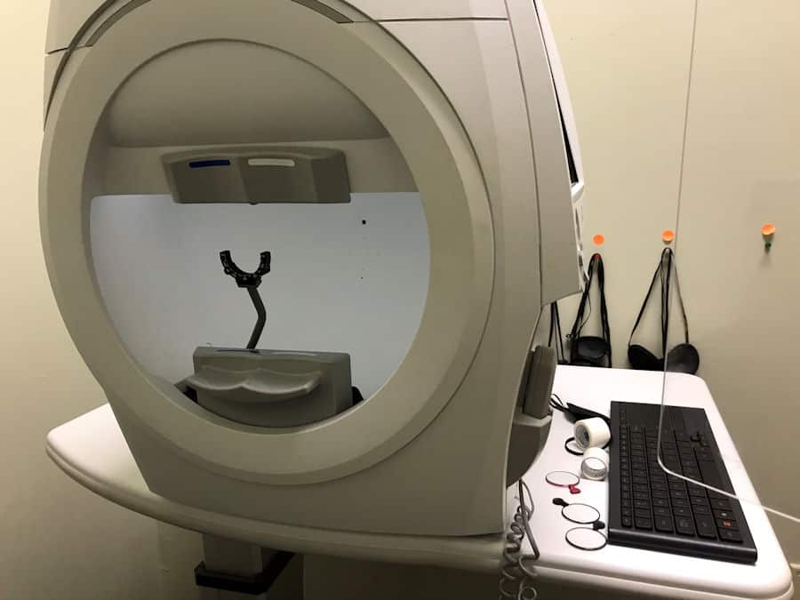
Humphrey Visual Field Instrument

Researchers have studied  Mirtogenol®, Ginkgo biloba, marijuana, baicalein, and forskolin for their anti-oxidant, anti-inflammatory, neuroprotective, or eye pressure lowering  effects, as natural glaucoma treatment alternatives to pharmaceutical drugs.

Glaucoma is a general term, used to describe numerous subgroups of the eye disease that result in optic nerve damage.  The simple definition is that it is a disease of high eye pressure, but it is actually more complicated than that.

The eye is constantly manufacturing fluid in the anterior chamber of the eye.  This fluid continually flows out of the eye by way of the blood stream.  Glaucoma is a problem of  reduced blood flow to the tissues of the eye. The decreased blood flow causes a  disruption in the flow of the fluid out of the eye. Fluid not filtered out of the eye causes an increase in intraocular pressure (IOP.)  Increased intraocular pressure (IOP) is a major risk factor for the damage to the nerves of the eye. This high eye pressure form of glaucoma is commonly called Primary Open Angle Glaucoma (POAG.)

But it is not the only risk factor.  Poor small blood vessel  circulation, especially at the level of the optic nerve and oxidative stres, are also risk factors. The type of glaucoma which is not characterized by high eye pressure is called Normal Tension Glaucoma (NTG.)

The damage of glaucoma is evident at the optic nerve, which is where the light-sensing retinal ganglion cell axons come together to form the optic nerve and exit the orbit of the eye on their way to the brain.  Death of these cells initially results in visual field loss, followed by loss of visual acuity.

 Glaucoma is chronic, progressive, lifelong, and potentially blinding if not treated.

The management and treatment goals of treating glaucoma are to slow the progression and stabilize the visual field loss.

In the early stages of glaucoma, the treatment of choice is  eye drops. While there are several different categories of medications, eye drops function in one of two ways:

* Decrease the production of aqueous fluid,  (ex: beta-blockers);  or

* Increase outflow of fluid, (ex: prostaglandins).

The taking of eye drops is lifelong and can be expensive.  Patients, very often, dislike taking eye drops.  It does not cure glaucoma. These treatments manage glaucoma.

Patients often ask if there is natural dietary supplements for glaucoma as an alternative to eye drops, lasers, and surgery.  Searching through the literature, I found 5 natural dietary supplements that claim to control glaucoma.  They are:

1.  Mirtogenol®: which is  Bilberry and French Maritime Pine Bark extract

2.Ginkgo biloba

3. Marijuana

4. Baicalein

5.  Forskolin

1. Eye Pressure Control: European bilberry +  French Maritime Pine=  Mitrogenol®

Mirtogenol is named for the combination of Mirtoselect® and Pyrogenol®.   Mirtoselect® is bilberry extract and Pyrogenol® is French maritime pine bark extract.

 Bilberry is a darkly pigmented berry found in northern Europe, which is high in anthocyanins.  Anthocyanins are flavonoids, which are the blue-black pigments of bilberry. These flavonoids have anti-oxidant and anti-inflammatory bioactivity.  Bilberry extract is also thought to have a neuroprotective (protects nerves from damage, degeneration, and impairment) function to enhance  the stability of retinal ganglion cells and the optic nerve. While it has many potential benefits, anti-oxidant, anti-inflammatory, and is neuroprotective, it does not decrease eye pressure. 

 Researchers are  also evaluating anthocyanins for their blood pressure lowering, blood lipid (LDL)  lowering, and blood sugar lowering effects.  Bilberry has long been associated with vision benefits.  Studies have shown that bilberry and vitamin E together have been successful in decreasing the progression of cataracts.  It has not been proven, by clinical studies, to increase night vision.

The extract called Mirtoselect® is a highly purified anthocyanin-rich extract, devoid of the non- anthocyanin components.

Pycnogenol® is a standardized plant-based extract obtained from the bark of the French maritime pine. This is a brand used by research studies because of its known purity.   It  has strong anti-oxidant, anti-inflammatory, and  capillary protective activity (this could be significant for those with diabetic retinopathy.)  Interestingly, the use of pine bark as an anti-inflammatory goes back to the time of Hippocrates.

Mirtogenol® is a combination supplement trademarked by Indena, Milan, Italy and Horphag Research, London, UK.

In a study , Mirtogenol® lowered IOP in those at risk for developing primary open angle glaucoma (POAG.) The mechanism is the combined activity of the two extracts to increase blood flow through the tiny blood vessels of the eye.  By increasing blood flow the inflow and outflow of fluid  stabilizes eye pressure.

Who should take Mirtogenol®?

Supplements with Mirtogenol® are indicated for those with elevated eye pressure and are at risk for developing glaucoma.

Dosage of Mirtogenol®

The dosage given in the study (referenced below) was Pycnogenol® 40 mg and Mirtoselect® 80 mg, twice a day.  The full eye pressure control effect  was reached at 3 months.  Reviewing commercially available ‘Eye Pressure Control’ supplements with Mirtogenol® lists the dosage at 120 mg (40mg + 80mg.)  The indication is to take with food to prevent gastric upset.

Safety

Both of these extracts, Mirtoselect® and Pycnogenol® have been used for decades as a dietary supplement without reported side effects.

Precaution recommends that Pycnogenol® is not for children under 6 years of age and  should not be taken during the first 3 months of pregnancy.

 (Ref: Effects of Mirtogenol® on ocular blood flow and intraocular hypertension in asymptomatic subjects

2. Ginkgo biloba as a Natural Treatment for Glaucoma

Ginkgo bil0ba are extracts from ginkgo biloba leaves that have been used for centuries in Asian medicine. The extracts are comprised of flavonoids and terpenoids.  It is credited to be an  anti-oxidant, anti-inflammatory, and neuroprotective (protects nerves from damage, degeneration, and impairment.)  Neuroprotection is  essential for those with  glaucoma, because it is the loss of retinal ganglion cells that results in vision loss. 

Ginkgo biloba is also effective in promoting small blood vessel circulation.  Gingko biloba  does not have an effect on eye pressure.  It is thought that it is this capability to increase ocular blood circulation  and its neuroprotective effect that  slows the progression of glaucoma.

Because of these bio-active effects, ginkgo biloba can be beneficial for prevention and treatment of other eye diseases such  as diabetic retinopathy, macular degeneration, and cataracts.

Who should be taking Ginkgo biloba?

Researchers have found that Ginkgo biloba  has potential to treat those with glaucoma caused by other risk factors besides high eye pressure.  (Remember, Ginkgo does not affect eye pressure.) This is significant for those with eye pressure in the normal range, but are still experiencing retinal ganglion cell damage (also known as Normal Tension Glaucoma.)  In these cases of progressive normal tension  glaucoma, it may be taken as a adjutant to medical therapy.

Recommended Dosage of Ginkgo biloba

Research studies most commonly used 40 mg extract, three times a day, for a total of 120 mg per day.   Reviewing commercially available supplements, the dosage given is 40 mg, 60 mg, or 120 mg. Extrapolating from research dosages, it would be 40 mg/3 times per day, or 60 mg/twice a day, or 120 mg/ once a day.

Safety of Ginkgo biloba

There have been reported incidents of spontaneous  bleeding for those at risk (risk factors like; other medications, old age, liver cirrhosis), who are simultaneously taking  non-steroidal anti-inflammatory drugs (NSAIDs. ex; hi-dose aspirin, ibuprofen, naproxen) and anticoagulants such as Plavix, heparin, or warfarin.  However, several studies could not implicate the combination treatment as the cause of bleeding.  Overall, Ginkgo biloba has a good safety profile.  It is always a good idea to discuss supplementation with your physician before launching into a new supplement regiment.

 (Ref: Ginkgo biloba: An adjuvant therapy for progressive normal and high tension glaucomaOpens in a new tab. )

3. Marijuana as a Natural Treatment for Glaucoma

Marijuana is the dried leaves and stems of the Cannabis sativa plant.  Historically, it has been used as a recreational  substance for its psychotropic effects.  It can be smoked or eaten (usually in baked goods), or drank as a tea. 

It’s significant extracts are ∆-9-tetrahydrocannabinol (THC) and cannabidiol (CBD).  It is the THC which is smoked, vaporized or taken orally for recreational use.  This active ingredient, over stimulates brain cell receptors. This is how the desired ‘high’ occurs for recreational users.

The mechanism by which marijuana lowers eye pressure is not fully understood.  Clinical data suggests that it may suppress aqueous fluid production and increase fluid outflow from the eye. It is also thought to have a neuroprotective effect.

THC based  medications ( Marinol) have been approved by the FDA as a treatment for nausea and appetite enhancement for cancer patients undergoing chemo-therapy and those with wasting diseases like AIDS.  Another extract, CBD  (there is an estimated 80 cannabinoids in marijuana) has been approved for a form of childhood epilepsy. THC and CBD (2 of the many cannabinoids ) combined with other medications  are formulated to treat spasticity and neuropathic pain. 

Safety and Side Effects of Treating Glaucoma with Marijuana

The marijuana plant has many other botanical compounds as a part of its structure.  It is these other compounds that can cause problems and adverse side effects when smoked or eaten.  

Short-term effects, besides the ‘high’ are; reduced blood pressure, increased heart rate, anxiety, and paranoia. Eyes become  red and dry. The pupils of the eyes become  small and light sensitive.

Consider, if the problem of blood flow to the eye is a risk factor for glaucoma, the reduction in blood pressure is counter to the benefit of lowering the eye pressure. 

Long term use (years) can affect thinking, memory, and learning capability.  Physically, frequent smoking of marijuana can irritate the lungs and increases the risk for substance abuse disorder.  It should not be used during pregnancy.  It has been credited for creating behavioral problems in these children. (Ref: Marijuana Drug FactsOpens in a new tab. )

Who Should Be Taking Marijuana to Treat Glaucoma?

Several studies have shown that the THC and other cannabinoids ‘significantly’ reduce eye pressure in both glaucoma patients and normal healthy individuals.

It was discovered 40 years ago that smoking marijuana decreased eye pressure (chief risk factor of glaucoma.) However, since that time, better therapies for treatment have been developed, which do not have the side effects of either smoking or ingesting marijuana.

At this time, there are no cannabinoid  medications approved for treatment of glaucoma.  The American Academy of Ophthalmology and the American Glaucoma Society do not recommend the use of marijuana to treat glaucoma.

Dosages of the Cannabinoids

The effectiveness of the cannabinoids is dependent on the dose, but is generally short-lived, 3 – 4 hours. Because it has a short-life of effectiveness, it must be taken frequently.

 This need to keep smoking or ingesting marijuana is not conducive to good ocular eye pressure control.  Contemporary  pharmaceutical treatments, which are effective without the side effects,  need to only  be taken once or twice a day.

The future of treating glaucoma with marijuana requires more clinical research to isolate the eye pressure lowering component (cannabinoids), which can be used as a glaucoma treatment without the adverse side effects. (Ref: Herbal Medicines in Glaucoma TreatmentOpens in a new tab. )

The next two herbal supplements are less well known as natural treatments for glaucoma:

 4. Baicalein

Baicalein  isolated from the Chinese medicinal herb, Scutellariae radix (Blue baikal Skullcap a.k.a. Chinese skullcap).  This active compound has been widely used in Asian countries for the treatment of many diseases.  It is a flavonoid compound and like most flavonoids, it has powerful anti-oxidant, anti-inflammatory, and neuroprotective activity.  (Flavonoids have also been proven to decrease cataracts and AMD.)

Pharmaceutical  eye drop treatment for glaucoma focuses on reducing eye pressure.  The studies on baicalein, thus far, have been animal studies (gerbils) and have shown that when introduced systemically, it reduced eye pressure.  Although research is ongoing, it is thought that baicalein reduces eye pressure  in two ways: 

1. decreasing  aqueous humor production at the ciliary body, and

2. it has been shown to facilitate the mechanism of outflow, leading to IOP reduction.  

It is anticipated that this supplement will be used as an adjuvant treatment for glaucoma in combination with standard glaucoma medications.

Safety of Baicalein

Baicalein has been used in traditional Chinese medicine with no recorded side effects.  Still little  has been done with safety and effectiveness in human clinical studies.   (Ref:   ARVOOpens in a new tab.)

5.  Forskolin

Forskolin is extracted from the root of the Indian Coleus, which is a plant from the mint family found all over India. Recently, it has gained fame as a dietary substance for weight loss.

The mechanism of action favorable for eye pressure control  is thought to be it’s ability to suppress the formation of aqueous fluid.

There are very few studies of forskolin as a treatment for glaucoma.  It was first studied in the 1980’s  when it was revealed to lower eye pressure in a small group of healthy patients.  The most significant  and hopeful study was one done in India.  Subjects used a 1% eye drop solution of forskolin three times a day.  The study indicated a 4 to 5 mm Hg average reduction in eye pressure. 

Other studies administered forskolin as an oral supplement combined with flavonoids.  This was less conclusive than the eye drop study because of the inclusion of flavonoids, which we know have anti-oxidant and anti-inflammatory, and are neuroprotective favorable for controlling glaucoma.  So the role of forskolin is less clear.

Safety of Forskolin

There is only limited information about any long term side effects.  Those who took the eye drops experienced some transient redness and/or irritation. 

Forskolin oral supplements have been shown to decrease heart rate and lower blood pressure.  So those taking blood pressure medications  should not take forskolin.  It is also recommended that those on anticoagulants and anti-platelet therapy should also  avoid forskolin.  There is evidence that it has androgenic activity on cells of the prostate and should be avoided by those with prostate cancer.

Dosage of Forskolin

Those in the Indian study administered 2 drops of 1% forskolin 3 times a day.  (This formulation is not available for purchase at the time of this writing.) The multiple of times (3 x per day) that the drops must be instilled detracts from its desirability as an alternative treatment.

Who Should Take Forskolin?

Forskolin has the potential to be a new class of glaucoma treatment.  The study out of India suggested that it may be a good alternative medication for those with asthma who cannot take beta-blockers (timolol.) ( Ref: Efficacy and safety of 1% forskolin eye dropsOpens in a new tab.…)

Right now, I would say to wait until more studies are done to prove its safety and effectiveness. 

In the End…

Keep in mind that the FDA evaluates and regulates prescription drugs and those drug treatments sold over-the-counter, but they do not regulate herbal medications and nutritional supplements.  For that reason, safety and efficacy studies are not required. Therefore,  herbal and nutritional supplements may  vary in purity and potency. 

None of the mentioned herbal supplements have the potential, at this time, to have the same efficacy as current glaucoma eye drop medications for lowering eye pressure. There is some potential for their neuroprotective effect  which reduces the loss of the retinal ganglion cells critical for  maintaining vision.  Most studies suggest that these supplements (predominantly Ginkgo biloba, bilberry, and Mirtognenol®)  should be considered as adjuvant to standard prescribed glaucoma medications.

The effectivity of these natural dietary supplements to control and treat glaucoma need larger and more in-depth studies to better gauge their potential. 

Can a Cup of Hot Tea a Day, Reduce the Risk for Developing  Glaucoma

tea pouring into a china teacup

The on-line British Journal of Ophthalmology published  a report indicating  that “Drinking a cup of hot tea at least once a day may be linked to a significantly lower risk of developing the serious eye condition, glaucoma.”

The investigators looked at an American study which yearly, surveys the lifestyle and health status of 10,000 Americans, by means of both physical exams and  interviews.

The researchers looked at the subjects consumption of coffee, tea, and soft drinks, both with caffeine and without caffeine.  After sorting through and teasing out the data (I won’t bore you with the numbers here),  the investigators determined that the drinking of a cup of hot tea with caffeine daily reduced the risk of developing glaucoma by 74%. In health research 74% is a BIG number. An almost Too-Good-to-be-True number.

So what is it?  Why would the caffeinated tea reduce the risk, but not the caffeine in coffee or soda?  For that matter, de-caffeinated tea was not a factor in reducing the risk, at least not in this study.  The American survey did not delve into types of tea (green tea, black tea,, Oolong tea, Lipton tea), brewing times, size of the cup, etc.  The information  was general: one cup (or more) daily.

Glaucoma is a progressive, sight threatening eye disease.  The common understanding is that it is a disease of high eye pressure, that damages the light sensing neurons of the retina of the eye.  The progressive  damage to these nerves results in nerve cell death, and subsequent loss in vision. Most therapies are centered on controlling the pressure inside of the eye. But doctors are finding that these therapies are sometimes not enough.  Medical research is focusing on other factors that contribute to the progression of glaucoma.

So that brings us back to the role that hot tea may play in reducing the risk of glaucoma.  The questionable aspect is the caffeine. Other research has shown that the eye pressure can go up, which is undesirable, when caffeine, in the form of coffee, is ingested. (Ref: JGlaucomaOpens in a new tab.)

Tea has other beneficial factors, such as antioxidants, anti-inflammatory, and neuro-protective substances.  These factors are also essential for good general health. They have been studied as an important nutritional component for heart and vascular health, diabetic control, and cancer prevention.  That which is good for the body is good for the eyes.

The researchers conclude that more research needs to be done to sort out the effects of tea on the risk for the development of glaucoma.

You may also be interested in…How Caffeine Affects Eye Disease and Causes Visual Disturbances
Astaxanthin and Vision Benefits


Flamingos

Astaxanthin is potentially the most powerful anti-oxidant and anti-inflammatory for the prevention and treatment of eye disease.

What are the health benefits of taking astaxanthin?

Astaxanthin is a nutrient of the carotenoid group. Similar to the other carotenoids, astaxanthin has anti-oxidant, anti-inflammatory, and immune stimulating effects. Studies indicate that it is the most powerful of the carotenoid anti-oxidants.

Although it has not been as thoroughly studied as other carotenoids, it has possible beneficial effects for cancer treatment, cardiovascular health, memory and brain function, skin anti-aging, and eye health.

Astaxanthin, lutein, and zeaxanthin are carotenoids that are not converted to vitamin A. This is important for those that are sensitive to the effects of vitamin A. (Stargardt’s disease)

It is important to understand astaxanthin bioactivity to understand how it can benefit eye health:

1. Anti-oxidant Activity

The carotenoids are important as a nutrient for their anti-oxidant capability to neutralize free radicals.  Free radicals  are unstable, molecules made by the process of cellular metabolism, that have the potential to damage the cells. When there is an excessive amount of free radical formation, the  potential for cellular damage is called  oxidative stress. Oxidative stress is the underlying cause of disease and aging.

One study was able to measure anti-oxidant activities among several other known anti-oxidants, astaxanthin, lycopene, lutein, vitamin E, α and β-carotene.  The study reported that astaxanthin was one of the highest performing anti-oxidants. (Ref: Antioxidant activities of astaxanthin..Opens in a new tab..)

Researchers have found that astaxanthin exhibits a unique anti-oxidant properties.  While most anti-oxidants perform their duties either on the inside of a cell or the outside of the cell membrane, astaxanthin has the unique capability to span the cell membrane to scavenge for free radicals on both sides.  This helps to maintain the integrity of that cell membrane. (Ref; Astaxanthin, cell membrane nutrient ...)

Light entering the eye, is a source for the formation of the damaging reactive oxygen species(ROS, which is free radical plus a non-free radical oxygen.) Because of its effective and specialized anti-oxidant capabilities, this supplement has important implications for those with eye disease, to decrease cellular damage and death.

2. Anti-inflammatory Activity

Inflammation is the body’s response to injury, infection, toxins, and radiation.  This response by the cells of the body is a beneficial defense mechanism that contributes to the healing process of the body. Sometimes, however, inflammation over-reacts and may become chronic.  This ongoing inflammation may not be the cause of a disease, but contributes to the disease process, rather than resolving it.  Examples of disease processes made worse by inflammation: inflammatory bowel disease, rheumatoid arthritis, asthma, diabetes, and uveitis.

The anti-inflammatory capability of astaxanthin is thought to be its ability to target those substances and enzymes produced by cellular inflammatory response.  It acts to inhibit and suppress formation of many inflammatory substances.

Ongoing research is looking at the anti-inflammatory benefits of  astaxanthin:

  • Neuro-degenerative diseases:  Alzheimer’s, Parkinson’s and diabetes mellitus
  • Chronic organ diseases: inflammatory bowel disease, inflammatory liver, and kidney disease
  • Chronic infectious diseases; H. pylori (gastrointestinal),
  • Anti-aging;  skin.

          (Ref: Astaxanthin and its Effect in Inflammatory.)

3. Immune-Enhancing Activity

Studies indicate that astaxanthin influences immune function.  It has been shown to increase immunoglobulin production (antibodies) and immune cell responses. In animal studies, it has been shown to increase T-cell and natural killer cell activity.  

Because of its immune enhancing capability, it is thought that it may play a role reducing immune suppression caused by UV radiation. This is significant in the study of skin cancers caused by sun UV exposure. (Ref: Astaxanthin in Skin Health…)

How Astaxanthin Benefits the Eye

The benefits of astaxanthin for eye health is still in the early stages, but recent data suggests that  it is effective in treating and preventing eye diseases.

Lutein and zeaxanthin are the two carotenoids studied in the second Age Related Eye Disease Study (AREDS 2). These two carotenoids comprise the macular pigment.

 Astaxanthin has been studied in smaller research projects, and not as extensively as these other carotenoids. It is not found to be concentrated in the macula. It may be as important to eye health as the other carotenoids because it has a more powerful anti-oxidant.

Several Eye diseases that may benefit from astaxanthin supplementation

Age-Related Macular Degeneration

The macula is the central area of the retina in the back of the eye that is responsible for our capability to see fine line and detail.  This area has a high demand for oxygen and energy. Because of its high metabolic activity, light exposure,  and oxygen use, it is subject to oxidative stress (lots of free radical formation.)

Oxidative stress and an accumulation of metabolic by- products results in an accumulation of drusen.  The lipofusin of the drusen is toxic to the supporting cells of the retina.  These cells, the retinal pigment epithelium (RPE), die off, along with the photoreceptors they support and results in subsequent vision loss.  This is the degeneration process of AMD.

Because astaxanthin is an anti-oxidant and has been shown to have UV absorbing capabilities, it is thought to benefit those with age related macular degeneration by reducing oxidative stress.

In another study, using mice,, it was shown that astaxanthin could suppress the growth of new, leaky blood vessels responsible for  accelerating wet macular degeneration.  (Ref; Inhibition of choroidal neovascularization…)

Concerned about Macular degeneration? See: How to Decrease the Risk of Age Related Eye Disease

Diabetic retinopathy

The retinas of those with diabetic retinopathy are subject to oxidative stress and inflammation.  Hyperglycemia in the tissues of the retina creates a cascade of glycation end products, resulting in oxidative stress and  blood vessel dysfunction.  If blood is not getting to the retina, ganglion cells of the retina begin to die.

Several animal studies have shown that astaxanthin reduces the oxidative stress and is neuroprotective.  It also acts to decrease the inflammatory cascade.  This activity can prevent the formation of choroidal neovascular nets(leaky blood vessels beneath the retina.)  These vascular growths of leaky blood vessels  are devastating to vision and the predominant cause of blindness for diabetics.

Glaucoma

Glaucoma is the result of elevated IOP, which leads to decreased blood flow to the retina. The decreased flow to the retina results in retinal ganglion cell death and loss of visual field and visual acuity. The treatments for glaucoma are targeted eye pressure lowering medications. 

Astaxanthin may play a role in protecting the nerves of the retina because of its anti-oxidant and anti-inflammatory capabilities

Cataracts

Normally, the lens of the eye is clear.  Cataract development is any opacity of the natural lens.  The most common cause of cataracts is aging, occurring after a lifetime of UV exposure, smoking, poor diet, and/or steroid use.  Diabetes and trauma to the eye can also result in cataracts.

 These events can cause oxidative stress to the eye and lens, resulting in changes to the protein matrix of the lens. The proteins are modified and aggregate, resulting in spots, vacuoles, clefts, and color changes..

Recent studies have shown that there is a delay in  development and prevention of cataracts,  when supplemented with astaxanthin because of its anti-oxidant activity..

Ocular Surface Disease

The surface of the eye is the cornea (clear) and conjunctiva (whites).  The front surface is subject to sunlight (UV exposure)and environmental assault (particles in the air), throughout our life.  

It is inflammation and oxidative stress that eventually cause a process called dry eye disease.  Symptoms range from uncomfortable to painful eyes.

The meibomian glands of the eyes, when inflamed produce little and poor quality tears.  With inadequate tears, the epithelium of the delicate tissues of the eye undergo oxidative stress and inflammation.

Studies done involving supplements for dry eye disease included astaxanthin among  other  anti-oxidants.   Supplementing with a combination of anti-oxidants for 8 to 16 weeks improved tear quality and stability and resolved corneal epithelial disruption.

Eye Strain

Eye strain or eye fatigue is a problem for those working each day on computer technology.  Those with low vision are especially troubled by eye fatigue, straining to use low vision devices.  Symptoms include; discomfort, headaches, pain, blur, and tearing.

 A study was done giving computer workers 5 mg once a day for 4 weeks.  The participants claimed “significant relief from eyestrain.”  In another study, a group in their 40’s experienced improvement in focusing at near.

Researchers thought that it is the relaxing effect on the internal ciliary muscle of the eye and increased blood flow to the retinal vessels that accounted for the improvement of eye fatigue symptoms. .  (Interestingly, the studies were all Japanese.)

What foods are high in astaxanthin?

Astaxanthin is made by small one cell microalgae, phytoplankton, yeast, and bacteria, Which is then a food source for small water creatures called  zooplankton and crustaceans  These micro-animals are consumed and the astaxanthin accumulate in  seafood including algae, shrimp, krill, lobster, asteroidean (sea stars), crustacean, trout,  and salmon.

Like many other carotenoids, astaxanthin is a pigment molecule. You can actually see the red color of astaxanthin in the pink shrimp, salmon, and lobster. Astaxanthin has been credited with making flamingoes pink, because of the shrimp they eat.

The Best Astaxanthin Supplements

Astaxanthin has  properties that suggest that it’s anti-oxidant potential may be the most potent among carotenoids.  It is not generally included in those vitamins that are labeled eye vitamins.

Because of the expense of naturally produced astaxanthin, the majority of supplements available are synthetically made from petrochemicals. The synthetic astaxanthin is used primarily for feeding marine animals to give them the desirable red-orange coloring.

Only a small part of commercial astaxanthin is extracted from natural sources. These are from cultivated algae and yeast, or other astaxanthin-producing biological organisms. Only the naturally extracted astaxanthin is approved for use in supplements.  You will note that on the supplement bottle it indicates alga Haematococcus pluvialis as the natural source, because  it is considered to have the highest capacity to accumulate astaxanthin.

How much astaxanthin should I take daily?

The Food and Drug Administration (FDA) has approved astaxanthin from the algae  H. pluvialis for supplementation dosages up to 12 mg per day and up to 24 mg per day for no more than 30 days. 

A quote from the European Food Safety Authority: (Feb. 2020)

“The Panel concludes that an intake of 8 mg ATX per day from food supplements is safe for adults even in combination with the high exposure estimate to ATX from the background diet.”

Most human trial studies used dosages of 4 to 12 mg per day.  Astaxanthin can be found commercially  as a lone supplement, but is thought to be most effective when combined with other nutrients.   

The best way to get astaxanthin is in its natural form by ingesting seafood. It is estimated that farmed Atlantic salmon was reported as 6–8 mg/kg flesh (2.2 lbs). For example, eating a 165 g (5.6 oz) salmon will provide an average of about 3.6  mg astaxanthin. (Ref: Astaxanthin: Sources…)

When to Take Astaxanthin

The carotenoids are lipids (fat soluble molecules.)  The absorption of astaxanthin is enhanced by the fat in the foods ingested. This suggests that astaxanthin is best taken with a meal containing  some dietary fats

What are the side effects of taking astaxanthin?

There is not much data implicating toxic side effects of astaxanthin.  The conclusion of numerous studies were that it is a safe nutraceutical. One study treated patients with 40 mg daily for 4 weeks with no apparent side effects. (A study on the effects against stomach H. pylori.)  This is considered research level of dosage.  Best to stay with FDA or European Medicines Agency recommendations.

The synthetic astaxanthin produced from petrochemicals has not been studied for safe human consumption. The  synthetic version has been shown to not be as effective as an anti-oxidant as astaxanthin naturally sourced. Supplements will and should be extracted from natural  sources such as microalgae,  Haematococcus pluvialis.  Watch for synthetic  astaxanthin to be labeled as ‘nature identical.’

The Eye Vitamins that include Astaxanthin

AREDS 2 Plus  by HealthHappiness  6 mg astaxanthin ((plus Vit C,E, zinc, copper, lutein, zeaxanthin, Fish oils (DHA,EPA)

Vision Alive Max by Holistic Health Labs  includes astaxanthin (plus anthocyanins,  lutein, zeaxanthin, and saffron.)

VisionMD by 1MD  4 mg astaxanthin plus lutein, zeazanthin, ‘mixed carotenoids,’ Vitamin E, and lycopene.)

OptiWell  byLiveWell  4 mg astaxanthin (plus lutein, zeazanthanin, Vitamin E)

Swanson ultra Vision Defense by Swanson  4 mg astaxanthin (plus lutein, zeaxanthin, bilberry, and ‘broccoli extract.’)

Liquid 20/20 Vision by Tropical Oasis  4 mg  astaxanthin (plus Vitamins A,C,E, zinc, lutein Zeaxanthin.)

All Natural Eye Support by Peak Performance  2mg astaxanthin (plus bilberry, lutein, zeaxanthin, and lutein.)

Eye Protector by Pure Synergy  515 mg carotenoids of which 2 mg is astaxanthin, 500 mg flavonoids, 50 mg enxymes.

Looking at this list, you can expect 2 to 4 mg of astaxanthin for these multi-supplements.  Only the AREDS 2 Plus by HealthHappiness had 6 mg.  Supplements that are 8 to 12 mg are usually sold as astaxanthin only.

Want to learn more, see our other article: 3 Most Commonly Recommended Supplements for Prevention of AMD

In the end… 

Astaxanthin is the most  powerful of the carotenoid anti-oxidants. We know that inflammation and oxidative stress underlie or are the cause of disease.  This potent anti-oxidant and anti-inflammatory activity has been demonstrated in many animal and human clinical trials.  Researchers study astaxanthin exploring potential roles for human health, both for treatment and prevention. 

A good reference:

Clinical Applications of Astaxanthin in the Treatment of Ocular Diseases: Emerging InsightsOpens in a new tab. Published 2020 may in Marine Drugs

Can Contact Lenses Help the Visually Impaired?


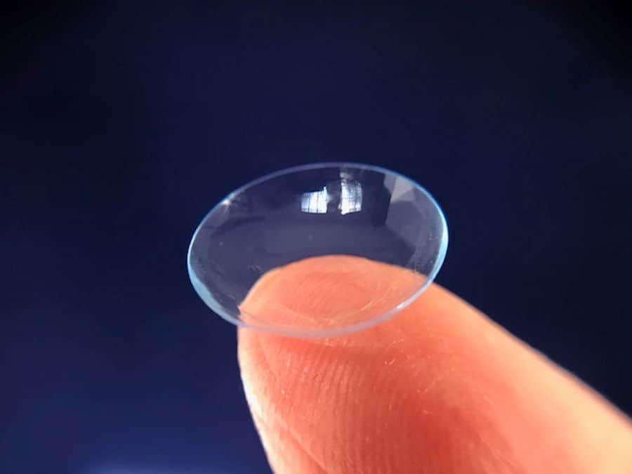
image of a contact lens perched on the tip of a finger

Contact lenses are used for those with low vision when there is:

  • High refractive error (otherwise, a thick eyeglass prescription),
  • therapeutic reasons: such as sensitivity to light and glare,  and nystagmus,
  • concerns about appearance:  to create a more normal look for increased  self-esteem, and
  • a need for either magnification or field enhancement.

1. Using contact lenses for high refractive error

How contact lenses are beneficial, compare to spectacle lenses:

  •  wider field of view,
  • wider field of fixation, because contacts move with the eye,
  • fewer oblique aberrations,
  • increased magnification for myopes (those who are nearsighted), and
  • contacts can help with eye discomfort relief when using low vision devices, which  can be held close to the eye to increase the usable field of view.

While correcting vision with contact lenses may not substantially improve distance visual acuity, especially for those with central vision loss, contacts do hold potential for enhancing vision in the peripheral areas of the retina. These peripheral areas may benefit from a sharper image.

Contact lenses work best for those low vision patients with high myopia, such as those who have pathological myopia.  That is because eyeglasses for those with high myopic correction , minifiy the visual field. In other words, the thickness of the eyeglass lenses makes  the world appear smaller.  Contact lenses, which rest up against the eye, do not create this high level of minification .  (Minification: the opposite of magnification.) In some cases, the use of the very thick eyeglasses may contribute to decreased visual acuity.

However, it’s worth noting that nearsighted individuals with near focusing problems or those who engage in a lot of close work may fare better with eyeglass correction.

2.   Therapeutic uses of contact lenses for those with low vision and eye disease

  • Tinted lenses reduce photosensitivity, glare, and
  • reduce eye movements (nystagmus).

Therapeutic Tinted  Contact Lenses

Photosensitivity  can be reduced or eliminated especially for those with albinism, cone dystrophies,  aniridia (those that lack an iris), and achromotopsia.

A small study reported that patients with retinal dystrophies benefited from a red-tinted contact lenses for the reduction of glare and an increase in contrast sensitivity.  (Contrast sensitivity is the ability to discriminate objects in an environment.) (Ref: : Red Tinted Contact LensesOpens in a new tab. )

For example: A child born with  achromotopsia(Congenital disease resulting in a severe  lack of color discrimination) lacks the ability to see color, is light sensitive, and has poor visual acuity.   These children benefited from a red/brown filtered lens which had the result of a slight increase in visual acuity and more comfort, even indoors.

For more information, here is an excellent article at Achromatopsia.infoOpens in a new tab.

Albinism and Therapeutic Contact Lenses

Those with oculocutaneous albinism may benefit by using contact lenses three ways:

  1. Tinted or opaque design lenses helps with glare and light sensitivity,
  2. Contact lenses are available in high prescriptions. (Reference to the above section on high refractive errors.)
  3. Contact lenses can also reduce the disruption of vision from the nystagmus, because the contact moves with the eye.

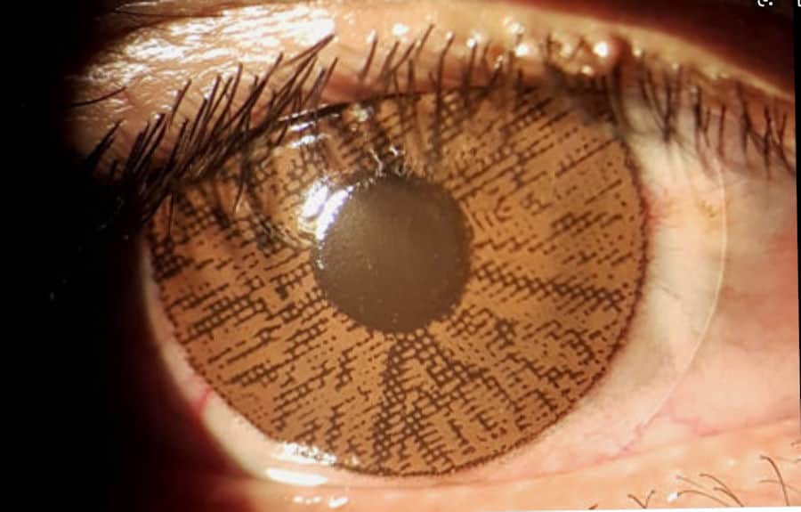
The most common type of specialty contacts prescribed for those with oculocutaneous albinism is a lens with an iris printed on and a clear pupillary center .  This lens design cuts down glare entering their very lightly pigmented eye. Case studies have shown that this contact lens with a printed-on iris can decrease light sensitivity. This type of lens is referred to as a prosthetic contact lens.

This printed-on iris also serves to give a more normal look to their eyes. 

Contact lenses are the preferred type of correction for nystagmus. Several studies have suggested that contact lenses can potentially reduce the frequency and amplitude of nystagmus, although this remains a topic of debate. This effect may be attributed to the correction of the high refractive error commonly associated with ocular albinism. Additionally, in certain patients, rigid lenses have been found to be more effective than soft lenses.

Contact Lenses to Reduce the Risk for Macular Degeneration and Cataracts

It is common knowledge that eye care professionals often recommend tinted eyeglasses for older patients to minimize UV exposure, aiming to reduce the risk of central retinal damage such as macular degeneration and cataract formation. Additionally, there are clear soft contact lenses available that incorporate UV protection into the lens material.

Commercially available lenses with UV protection:

All ACUVUE® Brand Contact Lenses have Class 1 or Class 2 UVblocking 

Biotrue One Day and Infuse 1 Day (Bausch + Lomb)  

ClearSight 1 day, Avaira(Class 1)/  Ray Ban 1 Day (Coopervision)

Biomedics 55 Evolution Aspheric and Biomedics 55 Premier Aspheric (Coopervision)

Each one of these manufacturer’s are careful to point out that there is no scientific data to prove that UVA/UVB contact lens protection will prevent macular degeneration or cataracts, and that wearing sunglasses outdoors is advised.

FDA Class 1 absorber. Recommended for high exposure environments such as mountains or beaches. The lenses in this classification must absorb more than:90% of UVA (316-380 nm wavelengths) and99% of UVB (280 – 315 nm)  
FDA Class 2 absorber. Recommended for general purposes. These lenses must absorb more than:  50% of UVA and95% of UVB  

Contact Lenses that Adjust to Different Light Levels for Light Sensitivity

Transitions® eyeglass lenses are the photosensitive plastic eyeglass lenses that change color to a darker ‘sunglass’ tint when exposed to UV light outdoors, and lighten up when entering  a space without UV light.   This technology has now been incorporated into Acuvue Oasys with Transitions® (Johnson & Johnson Vision Care) contact lenses. Like the eyeglass lenses, these contact lenses adapt to changes in  UV light intensity, filter blue light and provide UV protection.

They are also a useful option for computer and digital device users, because  the lenses adjust during use of technology that may be throwing off UV and high energy blue light.  The long term effects of chronic blue light generating technology on the health of our eyes is not yet known.

It is a good option for those with eye disease looking to reduce light sensitivity. Keep in mind, while this is an easy option for mitigating some UV exposure, sunglasses should still be worn outdoors. 

Contact Lenses that Deliver Medications

Contact lenses have taken on a new role beyond vision correction, now serving as a vehicle for the gradual release of medications over hours or even days. This innovative approach to eye care has made its debut in Canada and Japan with the introduction of the first medication-delivering contact lenses.

One such lens is designed to release an anti-itch medication, offering relief to those suffering from allergic conjunctivitis. By embedding the medication in a thin film along the outer rim of the contact lens, it avoids interfering with central vision. This not only provides targeted relief but also eliminates the need for the daily hassle and potential inaccuracies associated with traditional eye drop instillation.

Initially developed with a focus on a nighttime glaucoma medication known as latanoprost, these lenses pave the way for broader implications in eye care. This novel delivery system holds promise for addressing a range of eye conditions, including macular degeneration, diabetic retinopathy, allergy management, and post-surgical treatments.

Contact Lenses that Monitor Health

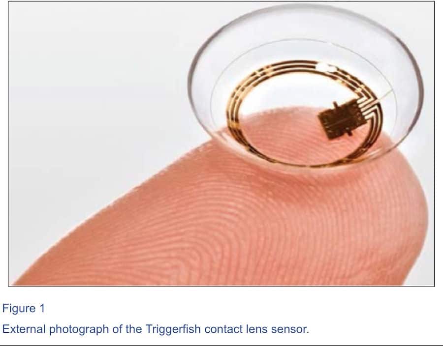
Recently approved here in the US is the Triggerfish® CLS (contact lens sensor) (Sensimed AG, Lausanne, Switzerland.)  This ‘smart’ contact lens is designed to detect eye pressure changes from the front surface of the eye over a 24 hour period.  It is designed to detect relative changes in eye pressure to help predict progression of damage due to glaucoma and determine affectivity of glaucoma treatments.

Interestingly, this small 14.1 mm contact lens has two gauges, a microprocessor and an antenna.  The eye pressure changes are relayed by the antenna to a portable recorder carried by the patient.

To read more click here: Sensimed’s Triggerfish


Contact Lenses that Monitor Body Fluids  

InWith Corporation along with contact lens manufacturer  Bausch + Lomb have created a prototype of a soft contact lens with microelectronics embedded in it to keep tabs on body chemistry, like blood sugar levels or to monitor other electronic body parts.  It will derive its energy from the natural blinking cycle of the eye and will send alerts and notifications to a smartphone.

See photos and video on InWith Corp. Facebook pageOpens in a new tab.

3. Enhancing Appearance and Self-esteem with Contact Lenses

For many individuals with visual impairments, the reliance on thick, heavy eyeglasses can sometimes amplify the appearance of their eyes, making them appear either significantly large or small behind the frames. Those with substantial eyeglass prescriptions might find themselves wearing what can feel like “coke-bottle” glasses, which, while aiding their vision, can impact their self-confidence.

Contact lenses, while not necessarily providing better vision than glasses, offer a sense of comfort and freedom for those who prefer not to be burdened by the weight and appearance of thick eyeglasses. Even though their vision might remain poor, the switch to contacts can make them feel less conspicuous and less encumbered by their visual impairment.

Furthermore, individuals who struggle with sensitivity to light and glare can discover relief by using-tinted lenses indoors and darker tinted sunglasses outdoors.

The choice to wear contact lenses can be transformative, especially for young individuals with visual disabilities, helping them feel more “normal” and socially accepted by blending in rather than standing out due to their eyewear.

4. How Contact Lenses are Used for Magnification and Field Enhancement

Telescopic contact lenses

This discussion is not about the head mounted or hand-held type of telescopes.  The two telescopic systems described here are  contact lens plus eyeglasses systems.  These are usually  calculated and set-up by an optometrists/low vision specialists.

The first system designed for magnification combines a high minus contact lens worn with a magnifying eyeglass lens.  This creates a ‘telescope’ lenses arrangement that can give up to 2X magnification.  This is a Galilean-style telescope for the optical geeks out there.  This contact lens/eyeglass system is best tolerated by younger persons with low vision. It can create some disorientation on head movement vs visual field movement.

How to increase the visual field for those with restricted visual fields; telescopic system:

Reverse contact/spectacle lenses telescopes to increase visual field for  those who have severely narrow visual fields, like those with retinitis pigmentosa. In order to increase the visual field, the contact lens is the magnifier(plus lens)  and the spectacle glasses (minus lenses) minifies the field so more can be seen.

These solutions utilizing telescopic systems to increase distance acuity or increase distance visual field can be disruptive to vision and will take time to adapt to.  As it is one eye that is set up for the telescopic vision and consequently disrupts binocular vision. 

At this time, these are the only contact lens-style magnification or field enhancing techniques.

But…there is new technology on the horizon:

Not yet available , researchers in California are developing a  contact lens that can zoom in to magnify and out to normal by using blinks. 

Another international group is developing a magnifying  contact lens that has  incorporated fitted mirror surfaces into a telescope a millimeter thick that fits within the contact lens.  This contact lens is combined with a set of polarized eyeglass lenses.

Both of these proposed technologies will benefit those who need magnification, especially those with central vision loss, like macular degeneration.

Considerations When Opting for Contact Lenses

Wearing contact lenses, whether for those with low vision or typical sighted individuals, comes with a set of responsibilities that should be carefully considered. Among these responsibilities are:

  • maintaining cleanliness,
  • proper handling, and
  • avoiding over-wearing contact lenses.

For visually impaired individuals, inserting and removing contact lenses can pose unique challenges due to the limitations of poor vision. While it is possible to do so by feel, the difficulty of finding a dropped lens can be a shared struggle, even for those with good eyesight. Additionally, the meticulous care required for cleaning and handling these small, delicate lenses can be daunting for someone with low vision.

The process of getting accustomed to inserting, removing, and caring for contact lenses is time-consuming and requires dedication. However, with sufficient motivation, it can be learned through touch.

It’s important to note that contact lenses prescribed to correct high refractive errors, similar to high prescription eyeglass lenses, tend to be thicker than average. This thickness can lead to discomfort, especially with extended wear.

Specialty lenses, such as those with printed iris rings, require precise fitting to ensure that the tinted ring does not interfere with vision. This often necessitates a fitting by a specialist and may involve a special order rather than an off-the-shelf purchase.

Finally, the issue of contact lens over-wear is a concern for many wearers, both sighted and visually impaired. Over-wearing occurs when lenses are left in for extended periods, often due to forgetfulness, convenience, or a strong preference for contacts over glasses.

Proper adherence to wear schedules provided by the prescribing doctor is crucial to avoid problems and complications. Neglecting these guidelines can lead to infections, redness, allergies, and eventually, an intolerance to contact lens wear. When considering contact lenses, it’s essential to weigh these factors carefully and be prepared for the commitment they require

Interested in learning more…Why those with low vision refused low vision aid

or Comparison of 10 Head-borne Digital Magnifiers for Low Vision

In the end…

It’s important for those with vision impairments to approach the idea of contact lenses with realistic expectations. Contact lenses may not necessarily improve visual acuity, but they offer a range of benefits discussed in this article. Whether it’s addressing light sensitivity and glare, opting for comfort over thick eyeglasses, enhancing appearance, or using specialty lenses for magnification or field enhancement, these are compelling reasons to consider contact lenses. By understanding these considerations, individuals can make informed decisions about whether contact lenses are the right choice for their unique needs and preferences.t.

How Vision Loss Affects the Social, Emotional, and Practical Aspects of Life


giant magnifying glass

Vision loss has profound effects on various aspects of life, influencing independence, self-perception, relationships, communication, economic status, expectations, and goals. Tasks that were once effortless now become challenging and time-consuming.

In our daily lives, the human eye effortlessly guides us through routine tasks with minimal forethought. Tasks such as personal care, medication management, reading, and household chores fall under the umbrella of activities of daily living. However, for those experiencing vision loss, these tasks must be approached differently, potentially leading to dependence on others for completion.

Fortunately, independence can often be preserved through training and a willingness to adapt by utilizing visual aids or adjusting living environments to suit individual needs.

Initially, acknowledging the need for assistance can be challenging, especially for individuals accustomed to a life with sight. While adjustments can be made to the living environment, there will inevitably be moments when external help is necessary. In these instances, it’s important to accept assistance to make challenging tasks more manageable.

Sometimes, well-intentioned family and friends may feel compelled to offer excessive help, inadvertently taking over tasks that the visually impaired person is capable of handling themselves. This can lead to a sense of helplessness and loss of control, ultimately increasing dependency.

It’s crucial to resist the temptation to allow others to take over tasks that can still be managed independently. Maintaining even a semblance of independence is pivotal in retaining control over various aspects of life.

For those facing more severe visual impairments, one of the most significant losses is the ability to drive. Suddenly, individuals find themselves reliant on others for transportation to run errands and attend appointments, which can be a difficult adjustment for those used to the freedom of coming and going as they please. This reliance, particularly in areas without accessible public transportation, often results in social isolation, bringing with it a host of negative emotions.

In cases of the most severe vision impairments, individuals may struggle with orientation and mobility. Orientation refers to knowing one’s location within an environment, while mobility entails the ability to move independently within that space. Fear of safety concerns such as getting lost or stranded may arise among the visually impaired.

To navigate these challenges, those with severe vision impairments or blindness often rely on tools such as canes or guide dogs to maintain their independence to the fullest extent possible

How Vision Impairment Affects Self-perception

It’s common for individuals to experience a decline in self-esteem and self-confidence. Independence may give way to dependence, and the loss of control over one’s life can create a sense of feeling ‘lesser’ than others, less valuable and capable.

The perception of this physical disability can significantly impact an individual’s sense of self-worth. They may begin to doubt their competence as a worker, parent, or spouse.

However, as those with vision loss begin to adapt and adjust, there is often a shift. Success in learning new skills and adapting to their circumstances can lessen the weight of the disability. Acquiring new abilities helps visually impaired individuals function better in their community and fosters the growth of self-confidence.

Relationships with family and friends are crucial for individuals who are visually impaired. Their understanding, acceptance, and willingness to assist play a pivotal role in the adjustment process for those with visual disabilities.

Navigating life without support can be significantly more challenging for visually impaired individuals compared to those with a supportive family environment. Friends and responsive healthcare providers can also offer the encouragement and support necessary for the individual to regain independence.

However, this support is not always guaranteed. In some cases, when a family member loses their vision, other family members may struggle to accept the resulting changes in family dynamics.

The roles within the family inevitably shift. Tasks that were once shared equally, such as household chores, errands, and driving, may now fall primarily on one or more family members to care for the visually impaired individual. These changes in responsibilities can also lead to shifts in financial burdens, potentially straining family members and household expenses.

Despite appearing “normal” and being able to manage many tasks competently, the visually impaired individual may face challenges if their family does not fully understand their impairment or denies that there is a problem.

This can create a situation where the individual hesitates to ask for help, as their family’s expectations have not adapted to their new circumstances. This added stress can be especially difficult for someone already grappling with anxiety and depression.

On the other hand, some family members may go to the other extreme, feeling the need to take over and handle tasks of any level of complexity for the visually impaired individual. They may hover, assisting with every step, which can inadvertently discourage independence and lead to a sense of incompetence in the visually impaired person. This dynamic can further erode their self-esteem.

Social situations present their own challenges. Friends and family members may not fully understand low vision, leading to misunderstandings and misinterpretations. The visually impaired individual may struggle to see details or recognize faces, potentially leading others to perceive them as unintelligent or clumsy. Additionally, in public settings like stores, clerks may become impatient with slower responses and movements.

Repeated incidents and accidents can further diminish social self-confidence and self-esteem for the visually impaired individual. This can create a cycle where they begin to avoid social interactions, ultimately leading to social isolation, increased depression, and anxiety

How Low Vision Affects Communication

Consider the myriad of places where printed information is found: on TV screens, product labels, signs, and even the numbers on appliance dials. Each of these can pose a challenge for individuals with print disabilities. (By the way, that includes those with dyslexia.)

The solution often involves enlarging (magnifying) the printed material, but it’s important to note that there isn’t a one-size-fits-all magnifier for every situation. The challenge for the visually impaired, caregivers, and therapists is to identify and utilize the most effective low vision aids, audio assistance tools, and environmental modifications. These aids are essential for enabling them to gather information and navigate their surroundings independently.

Fortunately, this generation of visually impaired individuals benefits from advancements in electronic and assistive technology. Developers of computer programs and smartphones have integrated assistive technology specifically designed to aid those with visual, hearing, and mobility impairments. However, it’s crucial that the visually impaired receive assistance with setting up and training to effectively use these technologies.

Despite these technological advances, one of the most challenging communication issues for the visually impaired remains the difficulty in distinguishing faces. The inability to recognize faces can lead to awkward social situations.

Human-to-human communication involves not just words and voice inflections, but also facial expressions that convey emotions and attitudes. These subtle non-verbal cues can be missed by the visually impaired, adding another layer of complexity to their interactions. Unfortunately, there is currently no technological aid that effectively addresses this aspect of communication.

How Vision Loss Changes Expectations and Goals

Parents of children with low vision hold hopes for their youngsters’ future education and physical development, much like any other parent would.

However, when low vision enters the picture, some of these plans may need to be set aside or adjusted. This adjustment often brings feelings of grief and sadness, particularly for those who experience sudden vision loss. On the other hand, individuals with slow, progressive vision loss typically have more time to come to terms with and adapt their expectations.

Seeking assistance from therapists and counselors can be invaluable in developing strategies for adjusting personal, work-related, and recreational goals.

Throughout this adjustment process, individuals often come to understand their limitations and develop new expectations. It’s crucial during this time to concentrate on abilities rather than disabilities. Channeling energy into learning skills that are both valued and enjoyable can be transformative.

Parents of visually disabled children are understandably concerned about the future their child will have. In the United States, government legislation such as the Rehabilitation Act of 1973, Section 504, guarantees educational options for children facing challenges in the classroom. Additionally, state and federal programs provide support until the age of 21. (Reference: Parents Information and Resources for a state-by-state listing.)

How Vision Loss Affects Economic Status

Fortunately, there are resources available to provide financial assistance to the visually impaired through government and state programs, particularly for educational and rehabilitation services. Medical care is typically covered by insurance, whether it’s private or through medical insurance exchanges.

However, it’s important to note that eyeglasses and low vision aids are often not covered by insurance. These costs are usually borne by the individual and their family. In the resources section of this site, I’ve listed a few groups that can help with locating and financing services and visual aids.

Click for Resources page.

In some cases, vision impairment doesn’t mean giving up one’s job. With the help of modern technology, disabled individuals can often continue working, depending on the nature of their job and the extent of their vision loss. Many individuals with low vision or blindness are able to remain gainfully employed.

The Americans with Disabilities Act (ADA) has established guidelines for employers with more than 15 employees. According to these guidelines, employers must make reasonable accommodations for employees with vision disabilities. In return, the employee must still be able to effectively and safely perform their job. For more information on these guidelines, you can Click VisionAwareOpens in a new tab..

Factors such as independence, self-perception, relationships, communication, and financial stability are all profoundly affected by vision impairment. If left unaddressed, these challenges can trigger a cascade of negative emotions.

Feelings of fear, anxiety, depression, loneliness, and helplessness often accompany the loss of vision, shaping the trajectory of life with low vision. These emotions are not uncommon among those who have experienced vision loss and can significantly impede the process of acceptance, adjustment, and adaptation.

The individuals who tend to fare best are those with a strong sense of self-esteem, coupled with a high degree of flexibility and a willingness to learn new skills and adaptations. These qualities are instrumental in navigating the complexities of life with low vision and achieving a successful and fulfilling existence despite the challenges.

In the End…

This journey typically involves a step-wise process of acceptance, adjustment, and adaptation. Successful individuals with low vision often learn compensatory and technical skills to aid in this adjustment.

The support of family, friends, and caring professionals can make a significant difference in helping the visually impaired regain self-assurance, self-esteem, confidence, and maintain independence. With time, patience, and the right support system, it is possible to navigate the challenges of low vision and continue to live a rewarding life.

How Caffeine Affects Eye Disease and Causes Visual Disturbances

Cup of Coffee at sunrise

 When it comes to eye health, researchers have found a link between caffeine and its impact on vision. Caffeine’s ability to block adenosine receptors in both the brain and retinal tissue plays a role in preventing inflammation, a destructive form of disease. This unique property of caffeine not only sheds light on its therapeutic potential but also suggests a beneficial effect on brain and retinal nerve tissue.

Further studies delve into the specifics of how caffeine interacts with our eyes. Research has shown that caffeine’s influence on adenosine receptors contributes to the dilation of blood vessels in the retina, leading to improved blood flow. This enhanced blood circulation is vital for maintaining optimal eye function and health.

Moreover, the antioxidants found in coffee, such as chlorogenic acid and melanoidins, have been linked to reduced risk of age-related macular degeneration (AMD) and cataracts. These antioxidants act as shields against oxidative stress, a major factor in eye diseases. It seems that a moderate intake of caffeine, through coffee or tea, could indeed be a boon for our eyesight in the long run.

Caffeine, Light Flashes, and Blurred Vision

Among the array of visual disturbances, the retinal migraine stands out as one of the most commonly reported, also known as an ocular, visual, or eye migraine. This type differs from the classic migraine, characterized by a visual aura followed by a migraine headache.

Signs and Symptoms of Retinal Migraine:

  • Area of blocked, grey or white vision with scintillating ‘broken shards of glass’ flashing lights.
  • Starts as a small area and seems to expand until finally dissipating.
  • Typically lasts from 5 to 20 minutes.
  • Often affects one eye, but often can’t tell if it is one or both eyes.
  • May occur multiple times in a single day.

The causes of ocular migraines remain a topic of debate. Most sources suggest it is attributed to a vasospasm of a retinal blood vessel. This reduction in blood flow to the retina can lead to the onset of light flashes and areas of transient vision loss.

Triggers for retinal migraines are varied, from emotional, physical, or chemical:

  • Caffeine: caffeinated drinks and chocolate,
  • alcohol,
  • stress, emotional and events like quitting smoking,
  • hypertension,
  • hormonal contraceptive pills,
  • hypoglycemia  (low blood sugar level),
  • changing to a higher altitude, and/or
  • exercise with overheating and dehydration.

Retinal (ocular, visual, eye) migraines can cause you to be alarmed. They have no known long-term consequences. If these occur frequently, consider lifestyle and dietary changes.  If making changes does not help, consider a medical evaluation for health-related concerns.

Caffeine and Eyelid Twitching (MyoKymia)

Frequent inquiries about eye twitching are a common occurrence in my practice. However, it’s important to clarify that technically, it’s not an issue with the eyes themselves, but rather the thin, delicate muscles of the eyelids that are responsible for the twitching.

Myokymia is characterized by the involuntary misfiring of nerves that innervate these fragile eyelid muscles. Individuals may experience episodes of eyelid twitching that persist for days or even months, with the exact causes often remaining a mystery.

Common triggers for myokymia include:

  • Caffeine consumption
  • Alcohol
  • Smoking
  • High emotional stress
  • Intense physical exertion and fatigue
  • Lack of adequate sleep.

Typically, myokymia does not impact vision significantly; it tends to be more of an annoyance than a serious problem. However, if other facial muscles or the deeper eye muscles responsible for eye movements become involved, a medical evaluation is necessary to rule out more serious nerve-related conditions.

Unfortunately, there is no specific treatment for eyelid twitching. However, it’s advisable to consider your dietary intake of caffeine, alcohol, and tobacco, as well as finding ways to reduce stress levels and ensure sufficient rest.

Caffeine and Eye Diseases

Cataracts and Caffeine

Our eyes, much like a camera, feature a clear natural lens positioned behind the pupil, akin to the aperture in a camera. This lens undergoes a natural aging process throughout our lives. In our younger years (under 40 years old), the lens is clear and flexible. However, as we age, it gradually stiffens and undergoes changes. Any alteration that affects the clarity of the lens is referred to as a cataract.

Signs indicating the presence of cataracts include:

  • Blurred or cloudy vision
  • Seeing halos around lights
  • Increased sensitivity to glare in sunlight
  • Difficulty seeing in dimly lit environments
  • Feeling like your glasses constantly need cleaning.

Globally, cataracts stand as one of the primary causes of vision impairment (according to the World Health Organization), often leading to cataract-induced blindness.

However, age isn’t the sole factor contributing to cataract development. Other factors include:

  • Nutritional deficiencies
  • Genetics
  • High exposure to UV radiation (particularly near the equator)
  • Diabetes
  • Smoking

In addition to the aging process, these factors can lead to the production of free radicals within the body. Free radicals are molecules generated through metabolic processes. Once formed, they can interact with other cells, potentially causing cellular damage. Such damage to the cells in the lenses of the eyes is a key factor in the development of cataracts.

Researchers have observed that populations consuming coffee exhibit a lower incidence of cataracts compared to those who do not consume coffee (Reference: :Effect of coffee (caffeine) against cataract blindness.”)

It is believed that caffeine and other antioxidants present in coffee work to reduce the oxidative stress caused by free radicals. Additionally, researchers have suggested the presence of other bioactive factors in coffee that contribute to its effectiveness in reducing the formation of cataracts. These beneficial substances are known as antioxidants.

Caffeine and Diabetic Eye Disease

Diabetes encompasses a group of chronic conditions that impact how the body metabolizes blood sugar, specifically glucose. When the body is unable to effectively utilize blood sugar, it leads to elevated levels of circulating glucose in the blood vessels. Over time, this can result in damage to the integrity of these blood vessels.

Diabetic retinopathy, blood leakage and macular edema

Within the eye, the result is diabetic retinopathy, in which blood leaks into the retina. Reduced blood perfusion means less oxygen is available to the retina, prompting the development of more fragile, leaky blood vessels (known as neovascularization) as a response to the low oxygen levels. Diabetic macular edema, characterized by fluid accumulation in the macula, is a significant cause of vision impairment in individuals with diabetic retinopathy. This condition arises from an inflammatory process within the eye.

The effects of caffeine and coffee on diabetic retinopathy are not extensively understood. However, a notable research study highlighted that caffeine can reduce blood vessel permeability. This effect is attributed to caffeine’s ability to block adenosine receptors.

In essence, caffeine blocks adenosine receptors in both the brain and retinal tissue, thereby potentially preventing inflammation. Coffee contains other active ingredients such as vitamins, minerals, and antioxidants. These additional properties of coffee may contribute to decreasing blood vessel leakage and mitigating the inflammatory processes associated with diabetic retinopathy.

Ref: Caffeine Prevents Blood Retinal Barrier Damage in a Model, In Vitro, of Diabetic Macular EdemaOpens in a new tab.

Caffeine and Glaucoma

Glaucoma serves as a broad term encompassing various subgroups of eye diseases that lead to optic nerve damage. While it is often simplified as a disease of high eye pressure, the reality is more complex.

This condition silently steals vision; aside from acute forms, there is typically no pain or noticeable visual disturbances.

The core issue in glaucoma lies in the outflow of aqueous fluid from the eye. The eye constantly produces fluid in the anterior chamber, which then flows out of the eye via the bloodstream. Glaucoma arises from a disruption in this fluid outflow process.

The impact of caffeine and coffee on glaucoma remains a topic of debate. Studies have shown a temporary increase in eye pressure following coffee consumption. This increase in pressure is influenced by the quantity of coffee consumed and an individual’s tolerance to caffeine.

The precise mechanism through which caffeine affects eye pressure is not fully elucidated. It is understood to raise blood pressure and constrict retinal blood vessels. Additionally, caffeine is known to increase the production of aqueous fluid in the eye, potentially affecting its outflow.

However, it has not been demonstrated to have an impact on glaucomatous damage in individuals already diagnosed with glaucoma.

A 2019 Korean study suggested a potential link between coffee consumption and an increased risk of open-angle glaucoma, particularly in men. Interestingly, this risk was not observed with tea or soda consumption. (Reference: “The relationship between coffee consumption and open angle glaucomaOpens in a new tab.“)

Research does not suggest that caffeine is a direct cause of glaucoma, nor has it been shown to prevent the condition.

Caffeine and Age-Related Macular Degeneration (AMD)

There is not a lot of studies on the effect of caffeine/coffee and AMD.  The best-known study that looked at the effect of coffee consumption and maculopathy was the Beaver Dam Study.

The Beaver Dam Study was a 15-year study of various aspects related to the development of maculopathy  of  3,917 persons, 43 to 86 years of age, living in Beaver Dam Wisconsin, starting in 1988.

Results as published by The Beaver Dam Study:

” Men were more likely to be coffee and caffeine drinkers than were women. For both men and women, coffee and caffeine intake decreased with age. Coffee and caffeine consumption was not associated with the 5-year incidence of early age-related maculopathy, soft indistinct drusen, or pigmentary abnormalities.”  Ref:  The relation of coffee and caffeine to the 5-year incidence of early age-related maculopathy: the Beaver Dam Eye StudyOpens in a new tab.

This study suggests that, based on its findings, there is no clear link between coffee or caffeine intake and the development of early age-related maculopathy, a common precursor to AMD.

In the End…

In exploring the intricate relationship between caffeine and the eye, researchers have highlighted a key aspect: caffeine’s influence on adenosine receptors. This mechanism is considered a therapeutic avenue, as it has the potential to prevent neuroinflammation, thereby protecting the nerves. Such findings carry significant implications for individuals with degenerative eye diseases.

Coffee, tea, and chocolate—common sources of caffeine—also contain a range of vitamins, minerals, and antioxidant compounds. These components may play a role in contributing to retinal health, potentially offering additional benefits beyond caffeine’s adenosine receptor-blocking properties.

As research continues to unravel the complexities of caffeine’s effects on eye health, it underscores the importance of moderation and balance in our consumption habits. While caffeine may present promising avenues for protecting the eyes and preserving vision, it is just one piece of the puzzle. A holistic approach to eye health, including a well-rounded diet rich in nutrients, regular eye exams, and a healthy lifestyle, remains crucial for maintaining optimal vision and preventing eye diseases.

“As long as one keeps searching, the answers come.” Joan Baez

Where to Get Help When You Have Vision Loss


picture of a girl wearing a headborne low vision device, looking outside.

Low vision services offer a multi-disciplinary approach to assist individuals with subnormal vision in accepting, adjusting, and adapting to maintain independence and quality of life. These services encompass a range of professionals including low vision specialists, rehabilitation experts, orientation and mobility specialists, teachers, and counselors.

If you’re seeking support for vision loss, these services can provide invaluable assistance tailored to your unique needs.

Who is a low vision patient?

Those who experience permanent vision loss, uncorrectable with eyeglasses or contact lenses, are candidates for specialized low vision services.

Individuals with vision less than 20/40 (6/12) in their better eye (where the other eye is worse) qualify for low vision services. Additionally, those who have experienced a loss of visual field (also known as “side vision”) or contrast sensitivity (difficulty distinguishing objects in similar shades of light and dark) can benefit from these services.

Referrals for low vision services are invaluable for individuals facing challenges with reading, driving, completing daily tasks, mobility, or experiencing emotional difficulties.

Low vision services are not only crucial for adults but also for children with inherited or acquired vision loss. Children have access to a range of therapies aimed at helping them adjust and adapt to their condition.

Who provides low vision services and where can low vision services be found?

Low vision services are available in various settings, including private practices (such as optometrists or ophthalmologists who specialize in low vision), multidisciplinary low vision centers, and state or federal agencies. These multidisciplinary centers often house low vision specialists who collaborate with occupational therapists, rehabilitation therapists, and counselors.

Low Vision Services:

The Starting Point:   Low Vision Evaluation

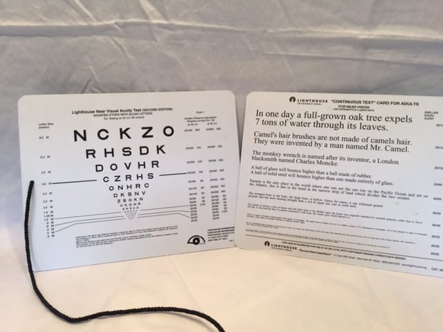
Low vision exam chart

Low vision specialists, typically licensed optometrists or ophthalmologists, possess a deep understanding of the disease process and prognosis due to their medical training. They are well-versed in functional vision loss and are aware of its limitations. These specialists have encountered individuals with similar visual challenges before, so they will not perceive age or debilitation as obstacles. Their primary objective is to assist in maximizing your visual potential and guide you through the adjustment and adaptation process.

Here are the reasons to get a low vision evaluation;

1. To Advocate for Yourself;

The low vision evaluation is your opportunity to interact with someone who understands your concerns can answer your questions.

2. Introduction to Visual Aids and Techniques;

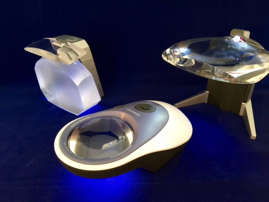
Optical magnifiers

The benefit of seeing a specialist is that he/she, because of knowledge and experience, can guide you in selecting the aids that would be helpful to you. 

3. Education and Counseling;

Prepare yourself for a very honest discussion about your impairment and options for rehabilitation.  The discussion should be tailored to you and your needs.

4. Appropriate Referrals;

They typically are familiar with state and local agencies for educational services, social services,  psychological counseling, and eligibility requirements.  They may also know of support groups for those with similar vision losses.

The low vision evaluation with a specialist commences with a comprehensive history and a series of questions to discern your concerns and requirements. The aim is to enhance your abilities in education, work, self-help, and recreational activities.

The tools utilized by the specialist may differ slightly from those used by your regular optometrist or ophthalmologist. Charts and lenses are employed to assess your level of visual function, determining the amount of usable vision and how effectively you can utilize it.

image of low vision charts used by low vision examiners
Low vision charats

Subsequently, the specialist will introduce various visual aids, including both optical and non-optical options, and recommend techniques to optimize your vision. Optical aids encompass items such as magnifiers and digital technologies, while non-optical aids include objects with large print (such as phones and kitchen items) or improved task lighting. Visual techniques involve methods like eccentric fixation and scanning.

After identifying the optical and non-optical aids that best suit your needs, the specialist can refer you to an expert who will assist you in utilizing these aids effectively and implementing low vision techniques.

Rehabilitation Services

Rehabilitation services help to identify and train you in strategies and techniques useful to maximize remaining functional vision.  Adaption is strengthened by training and use of compensatory skills. 

Areas of Rehabilitative Training:

CCTV desk top magnifiers

1.  Work.  An occupational therapist will help modify the work environment and train in the use of optical and non-optical aids used for meaningful and purposeful employment.

2. Home management.  The therapist will modify and train in the use of adaptive independent living skills.  This may require a visit by the therapist to the home. They can help by modifying the lighting, arrangement of furniture, and setting up optical aids.  Kitchen modification include not only optimal lighting , but contrast enhancement, large print kitchen objects, and tactile clues.

3. Personal care.  The therapist can help with organizing and labeling of personal care items in the bathroom.  This may also include the organization and labeling of medications for accurate access. There is magnifying mirrors and magnifying electronics to help with visualizing hair grooming and makeup application.

4. Communication.  There are large button phones and phone services available.  Especially interesting in this area are the artificial intelligence (AI) devices that respond to voice commands and can perform internet access tasks.

5.  Community access.  The therapist can advise and help the low vision person with accessing transportation and local services.

Navigating the World with Orientation and Mobility Training

For those with more severe impairment, the guidance of an Orientation and Mobility Specialist becomes invaluable. Orientation and mobility training focus on fostering independence and safety, aiding individuals in navigating their homes and public spaces.

Assistive devices used for mobility may include electronic aids, canes, guide dogs, and other walking aids. These specialists teach visually impaired individuals to utilize their senses and any remaining vision, as well as the proper use of a cane.

woman with raincoat and boots, using a cane for mobility

Certified Orientation and Mobility Specialists provide strategies for indoor and outdoor movement. They assess homes and workplaces for safety and ease of movement, using this information to assist individuals in orienting themselves and moving successfully. This training extends to navigating urban areas like streets and public transportation.

The skills imparted by an orientation and mobility specialist greatly enhance the confidence, independence, and safety of visually impaired individuals.

Supporting Mental Well-Being: Counseling Services

Psychiatric and social counseling services are not universally available in every low vision center. These services fall within the domain of psychiatrists, psychologists, and social workers. When confronted with psychological and social challenges, individuals with visual impairments may silently struggle. Doctors, family members, and friends might not immediately recognize this need.

The complexities of vision loss can be compounded by additional health concerns or challenging family and living situations. Those grappling with such difficulties may benefit from counseling services beyond what rehabilitation therapists typically provide.

Research indicates a correlation between the degree of vision loss and the prevalence of depression. Individuals with greater visual impairment often experience more pronounced depressive symptoms.

Conversely, studies have shown that those who received  low vision services had a reduced level of depressive symptoms. ( Low Vision Depression Prevention ….)

Remember to advocate for yourself.  Vision loss can be a very painful and emotional mine field.

Want to learn more, see the article  Low Vision, Depression, and AnxietyOpens in a new tab.

Low Vision Services for Children: A Comprehensive Approach

Children and teenagers may experience inherited forms of low vision or acquire them during their early years, encompassing their first and second decades of life. This includes conditions such as pediatric cataracts, pediatric glaucoma, nystagmus, and inherited retinal dystrophies.

The extent of low vision services offered will vary based on the child’s age at diagnosis. Concerns for an infant will differ from those of a child already attending school.

In the first year or two of life, the focus is on the child’s brain development and functional vision. For school-age children, the emphasis shifts towards literacy development and adaptive skills.

Rehabilitation therapists and teachers specializing in low vision for children possess expertise in this unique area of care. They are well-versed in the vision development of children and specialize in tailoring therapies to each child’s needs.

These specialists collaborate not only with the student and family but also with other members of the child’s educational team. Together, they establish a personalized plan for instruction and implementation.

The significance of early diagnosis and treatment, both medically and for vision rehabilitation in young children, cannot be overstated. The educational and adaptive support provided to school-age children is pivotal to their future. With proper intervention, their future can be just as rich and productive as that of their sighted peers.

For those parents not sure about the rights of their disabled child, see section 504 of the Rehabilitation Act.  There are many good websites explaining  this document, click for The U.S. Department of EducationOpens in a new tab..

The Role of Teachers of the Visually Impaired

Teachers of the visually impaired play a crucial role by integrating the diagnosis with information from other specialists and therapists, conducting their own functional assessments within the school setting. They become advocates for the students, interpreting medical information and comprehending the unique needs of each low vision student. This information is then communicated to the educational team, ensuring that the student has appropriate access to academic learning.

When to Consider Referring for Low Vision Services

Based on the criteria outlined earlier, it is generally recommended to refer individuals for low vision services when their visual acuity is measured at less than 20/40 (6/12), or when there is visual field loss or loss of contrast sensitivity, as diagnosed during the eye examination by your eye doctor.

Here is a better determinant of low vision  by the National Eye Institute:

 “Low vision is a vision problem that makes it hard to do everyday activities. It can’t be fixed with glasses, contact lenses or other standard treatments like medicine or surgery.”

In other words, if vision loss is affecting your lifestyle, activities, work, and happiness, it’s time for a referral for low vision services.

Understanding Functional Vision

There is a growing movement to reassess a patient’s functional vision not solely based on numerical measurements like visual acuity, degree of field loss, or contrast sensitivity, but on how these factors affect the individual’s daily functioning. These assessments are conducted by low vision specialists. While these numbers provide valuable data, they may not fully capture the individual’s actual visual experience, which can vary widely among people with vision impairments.

For instance, an individual with a visual acuity of 20/40 might be mildly bothered by the decrease in clarity but find it does not significantly impact their work or hobbies. Conversely, an artist or editor may experience profound despair due to the loss of visual detail at the same acuity level.

Consider someone with glaucoma who has lost a sector of their vision. The extent of impairment will be influenced by the size and location of this visual field loss.

Another example is a person with decreased contrast sensitivity, often due to cataracts. One individual might hardly notice the loss, while another may feel a constant unease due to the perceived dimness in their surroundings. It’s possible to have poor contrast sensitivity yet maintain good visual acuity.

Additionally, individuals with conditions like macular degeneration or Stargardt’s disease may have complete central vision loss, leading to a “legally blind” classification. However, with appropriate low vision aids and techniques like eccentric fixation, they can function effectively in daily life.

It  is  how you use your remaining vision and how you function with your vision loss:

  • Compensatory techniques, with and without aids,
  • adaptions to surroundings,
  • visual skills, like eccentric viewing and scanning,
  • add in; use of other senses, like hearing and touch.

Specialists in low vision, rehabilitation, mobility, and counseling conduct comprehensive functional vision assessments. These assessments encompass inquiries into how vision loss has impacted various aspects of your life, including daily activities, work, personal needs, and psychological well-being.

The assessment also involves tests to gauge your progress in developing visual compensatory skills and adaptations. The results of these assessments guide the specialists in formulating personalized plans for low vision services tailored to your specific functional vision needs.

What problems do you identify:

  • 1.Reading and writing,
  • 2. seeing the television,
  • 3. unable to see the screens of  computer or other electronic devices you need,
  • 4. difficulty with recognizing faces,
  • 5. unable to identify dials on household appliances,
  • 6. headaches and eyestrain trying to see,
  • 7. find yourself bumping into corners , missing stairs, or curbs,
  • 8.  persistent feelings of sadness or anxious,
  • 9. spending more time alone?

In the End..

Specialists in low vision, rehabilitation, mobility, and counseling conduct thorough functional vision assessments. These evaluations delve into the impact of vision loss on your daily activities, work, personal needs, and psychological well-being.

Through these assessments, specialists can gauge your progress in developing visual compensatory skills and adaptations. The outcomes of these evaluations serve as the foundation for crafting personalized plans for low vision services. These plans are uniquely tailored to address your individual functional vision needs, providing a roadmap to enhance your quality of life and independence.

A study at a VA setting showed that those who received rehabilitation therapy along with low vision devices did better than those who only received low vision devices. Reference: (Veterans Affairs Low Vision Intervention Trial II Opens in a new tab.)

If we did all the things we are capable of doing, we would literally astound ourselves. Thomas Edison