Insight into Low Vision

Recent Posts

Pretending Not to be Visually Impaired: Passing with a Disability


black and white close-up of a pair of eyes looking upward

Individuals with vision impairment often adopt behaviors and tactics to hide their disability, striving to blend in and appear “normal.” This is facilitated by the fact that vision impairment is often categorized as an “invisible” condition.

What is Passing?

“Passing” in the context of disability refers to the intentional act of concealing one’s disability. It involves individuals with disabilities attempting to appear as if they do not have a disability, especially in social situations. This can involve hiding behaviors or characteristics associated with their disability to blend in with able-bodied individuals.

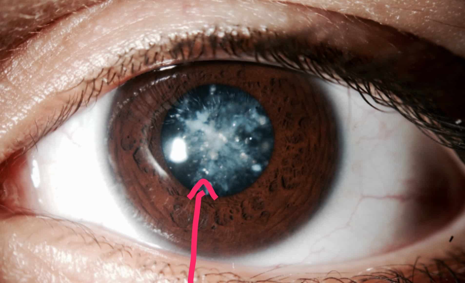
Vision impairment is often considered “invisible” because it may not be immediately apparent to others when interacting with someone with this disability. There are typically no outward signs of vision loss unless a person exhibits specific low vision behaviors, such as holding objects very close, tilting their head, excessive blinking, or eye movements that appear unfocused or exaggerated.

Passing may seem deceptive or dishonest to some, as individuals are essentially pretending to be able-bodied when they are not. However, for many with vision impairment, the decision to pass as fully sighted is deeply personal and often driven by emotional reasons.

Living in a world between the blind and the sighted, those with visual impairment often use passing as a form of self-defense. It allows them to create a façade of normalcy, helping to protect their social acceptance and preserve their psychological well-being.

Why the visually impaired conceal their disability.

Social Pressure: The social stigma attached to the term “disability” can subconsciously compel those with invisible disabilities to conceal their condition, aiming to sidestep the negative social implications of deviating from what is considered “normal.”

Lack of Understanding: Many individuals without visual impairments may not fully comprehend what “low vision” entails. Social norms often equate disability with a perceived lack of capability, extending beyond just the ability to see. Vision impairment is sometimes mistakenly equated with blindness or even intellectual disability.

They may find it is just easier to pretend. Openly identifying themselves as disabled, means opening up the “I can’t see, but I’m not blind” litany.

Ease of Passing: At times, “passing” as sighted is simply easier than delving into detailed explanations of personal limitations. This approach helps avoid the use of terms like “disabled” or “impaired,” which can steer conversations into uncomfortable or negative territory. Responding to such terms, listeners may exhibit patronizing attitudes or expressions of pity. To sidestep these reactions, visually impaired individuals may choose to pass as sighted for their own comfort and that of those around them.

Persistent Biases: Despite advancements in legislation and changing social attitudes toward disabilities, personal biases still linger, affecting how individuals perceive and react to disability. These lingering biases contribute to the inclination to conceal one’s visual impairment in social settings.

Self-Image: For those newly experiencing visual impairment, acceptance of their low vision may still be an ongoing journey. Choosing to remain silent about their disability can be a defense mechanism to uphold feelings of self-worth and a sense of “normalcy.”

Social Stigma and Self-Esteem: The societal stigma surrounding disabilities can lead to negative self-perceptions among the visually impaired. They may struggle with feelings of low self-esteem, perceiving themselves as “less-than” others.

Our self-perception is often influenced by how others perceive us. By not openly declaring “I am visually impaired,” individuals with disabilities aim to maintain equal standing with their peers. This is an attempt to resist being solely defined by their disability.

Privacy Concerns: Disclosing one’s disability and sharing personal details can feel invasive. Many visually impaired individuals view their visual status as private information, to be disclosed on a need-to-know basis.

Shame and Embarrassment: Past experiences with impaired vision that led to embarrassment or feelings of inferiority can result in a deep sense of shame. Others seem superior because they can see more quickly, react appropriately, and are queued into nuances of social interaction.

All of these seemingly small details of social interaction work due to better visual acuity. Facial recognition, facial expression, and visual jokes are missed by those with low vision leaving them to feel left out and embarrassed. Passing as sighted becomes a method of hiding what they perceive as a weakness or inadequacy.

Social Exclusion and Fear: Fear plays a significant role in this dynamic:

  • Fear of loss of social acceptance.
  • Fear of being marginalized or excluded and loss of social status.
  • Fear of losing a job because of assumptions made by others who do not understand .
  • Fear of Self-Realization: Admitting to oneself that they are not “normal” can be a daunting and fearful prospect.
  • Fear of Social Judgment: The anxiety of having to explain their visual impairment in detail, knowing others will pass judgment.
  • Fear of being singled out or treated differently due to their disability.

In navigating these complex emotions and societal pressures, visually impaired individuals often find themselves balancing between maintaining their privacy, preserving their self-image, and navigating a world that often fails to fully understand their experiences.

Also see this article on Why Those with Low Vision Have a Problem with Facial Recognition

How Children Learn to Pass

Pretending to See: Children often adjust their behavior to match what they believe others expect of them, sometimes pretending to see more or less than they actually can.

Parental Influence: Children are keen observers of their parents’ attitudes and fears. They quickly pick up on the concern surrounding their visual impairment, learning that not seeing what others see is viewed as a problem. To gain approval and avoid worry, children may give false responses, pretending to see things they actually can’t. The parent’s relief and positive reinforcement become a reward for their pretense.

Avoidance and Ignorance: Parents may sometimes choose to ignore the issue, perhaps due to difficulty in facing the reality of having a visually impaired child. This avoidance communicates to the child that their visual disability is something to be disregarded, contributing to their learning to “pass.”

Negative Attention in School: As school-age children, they quickly learn that being different can attract negative attention from peers. Other children may not always be kind or understanding, leading to feelings of exclusion and loneliness.

Treatment and Bullying: Disabled children may be treated as “special,” often being segregated from other children and excluded from certain activities due to accessibility challenges. They might also be infantilized, treated more childishly than their sighted peers. This difference in treatment can make them targets for bullying, which can be deeply upsetting and isolating.

Academic Impact: In attempting to pass as normal and avoid negative attention, children may also avoid asking for necessary help. This can result in falling behind academically, as their reluctance to seek assistance hampers their learning progress.

These experiences of social exclusion, bullying, and the pressure to conform to societal norms can have profound effects on a child’s development and self-esteem. As they navigate these challenges, children with visual disabilities often learn to hide their disability as a means of coping with the complexities of their social environment.

The consequences of pretending to be normal.

Social Awkwardness: When others are unaware of the limitations faced by someone concealing their visual impairment, the visually impaired individual may appear socially awkward. Due to their low vision, they might struggle to recognize faces, interpret facial expressions, and grasp nonverbal cues essential for social interaction. This can lead them to appear distracted or unresponsive in social situations.

Discomfort and Embarrassment: Admitting to not being able to see adequately after concealing it for some time can be uncomfortable and embarrassing. Those who were unaware may feel suspicious upon learning this information, wondering why it was kept hidden.

Navigating Social Situations: Navigating social situations with a visual impairment is inherently challenging. This challenge is compounded when others fail to understand the social mistakes that might occur, mistakes not typically made by those with normal vision. Consequently, visually impaired individuals may withdraw from social interactions, leading to feelings of depression and anxiety.

See my article on: Low Vision, Depression, and Anxiety

Employment Challenges: Concealing visual impairment from potential employers can pose significant challenges. Passing as sighted may seem like a way to bypass personal biases that some employers have against hiring disabled individuals. Despite legislation aimed at preventing discrimination, it unfortunately still exists.

Lack of Reasonable Accommodations: Failing to disclose vision loss to an employer means that necessary adjustments to accommodate low vision won’t be made. This can hinder the visually impaired individual from performing their job to the best of their ability. While some employers may be willing to make these adjustments, others may not understand or be open to the idea.

Risk of Errors and Job Loss: Attempting to pass as sighted comes with the risk of making unnecessary errors due to unmet visual needs. This could potentially result in losing a job for which reasonable accommodations could have been made.

Opportunity for Education and Awareness: Revealing one’s limitations is not just about personal acceptance; it’s also an opportunity to educate others. Just as the LGBTQ community has brought awareness and acceptance to their experiences, those with disabilities can also benefit from greater understanding. It’s crucial for others to realize that individuals with impairments are simply a different form of normal, deserving of respect, understanding, and the accommodations necessary to thrive.

In the end…..

Passing with a visual impairment presents numerous challenges, from social awkwardness to employment uncertainties. The decision to conceal one’s disability is often driven by social pressures and fear of stigma. However, there can be consequences leading to discomfort, social difficulties, and missed opportunities for accommodations.

Revealing one’s limitations can promote understanding and empathy, fostering a society that values inclusivity. By supporting individuals with visual impairments, we can create a world where everyone can thrive.

Read more about the Experience of Being Visually Disabled: How Vision Loss Affects the Social, Emotional, and Practical Aspects of Life

Why Those with Low Vision Have a Problem with Facial Recognition


featured image, set of graphic faces

Individuals coping with low vision face challenges in visual acuity and stable fixation, making facial recognition particularly difficult. This crucial skill relies on the ability to perceive detail and maintain steady focus for the brain to interpret the visual input.

Recognizing faces is fundamental for successful human interaction, yet it poses a significant hurdle for those with central vision loss. Conditions such as macular degeneration, Stargardt’s disease, and various cone-rod dystrophies are commonly associated with difficulties in facial recognition.

Two Factors Contributing to Decreased Facial Recognition Ability

1.  Eccentric viewing. In advanced stages of central vision loss, individuals often rely on a technique known as eccentric viewing, where they use parts of their retina outside the central macula. Eccentric viewing areas are the regions surrounding the central vision area, enabling those with vision impairment to “push” the blind central area aside and focus on objects using these alternative areas. However, due to how the retina is wired to the brain, these peripheral eccentric viewing areas struggle to maintain stable fixation, especially when attempting to focus on a face.

This is a departure from the normal central vision area, which can efficiently lock focus onto a face. The stable fixation in the central vision area allows the brain to more effectively analyze the facial image and make connections to areas of memory.

(Note:  This area is often referred to as the preferred retinal locus (PRL), meaning, the visually impaired has learned to use this other area of the retina as the area they prefer to use, rather than the ‘blind’ central vision area.)

Ref: Abnormal Fixation in Individuals With Age-Related Macular Degeneration When Viewing an Image of a FaceOpens in a new tab. Optometry and Vision Science.

This leads to the second factor affecting the capability of those with low vision to recognize faces;

2. Acuity.  When individuals with low vision use a peripheral viewing area of the retina (PRL) or rely on a blurry central vision area (macula) to gather visual information, the brain struggles to discern the fine details crucial for facial recognition. Unlike the central vision area, the eccentric viewing area lacks the high density of cone neurons necessary for precise line and detail discrimination defined as good acuity.

Facial recognition hinges on the ability to perceive the subtle nuances of different facial structures. However, the brain receives an unstable image due to the lack of finer details, poor visual acuity, making accurate facial recognition challenging.

In an attempt to compensate, individuals with low vision focus on the details they can perceive, often relying on auditory cues as well. They may observe general features such as the shape of the hair, face outline, and height.

For instance, a person with low vision might recognize that an individual has eyes, a nose, and a mouth, but the distinguishing features are unclear and therefore difficult to remember. The unique characteristics of the eyes, nose, and mouth, including their structure and proportions, which are crucial for differentiation, remain obscured.

The Challenge of Facial Expression Interpretation for the Visually Impaired

Facial expressions is a form of nonverbal communication, conveying emotions and signals integral to conversations. The interpretation of facial expressions occurs rapidly and unconsciously, allowing individuals to detect a wide range of emotions and personality traits that may not be apparent from tone of voice or spoken words alone.

Expressions such as happiness, sadness, scorn, fear, surprise, distraction, anger, and various personality characteristics are conveyed through facial cues, enriching the meaning of a conversation and conveying emotions to the listener.

In typical conversations, listeners often mirror the emotions and feelings expressed by the speaker, creating a natural flow of understanding. However, individuals who are visually impaired lack this additional layer of information conveyed through facial expressions. This absence places them at a disadvantage in comprehending and responding appropriately to the emotional context of a conversation.

For those with visual impairment, understanding is primarily reliant on verbal cues such as volume and tone of voice. The inability to “read” facial expressions can lead to misunderstandings where a visually impaired individual may seem inattentive or dismissive of the speaker’s emotions or intentions.

Consequently, relying solely on verbal cues can make the visually impaired individual feel ill at ease, anxious, or embarrassed in social interactions. It is not uncommon for individuals with low vision to avoid social situations due to the challenges posed by the inability to interpret facial expressions, which are so integral to human communication.

 Ref; Role of facial expressions in social interactionsOpens in a new tab.

The case for emojis:  Think about reading a text message and being unsure of the writer’s intent. You might wonder, ‘Are they angry, joking, sarcastic, or annoyed?’ There is no facial expression or tone of voice to gauge what the writer is conveying.  This is the ‘lost’ feeling those with vision impairment feel in some social situations.  Luckily for the individual who is texting, they can add in an emoji to clarify their feelings!

The Challenge of Interpreting Facial Expressions for the Visually Impaired

Social embarrassment

Being able to see and recognize a face imparts more than just the speaker’s feelings and emotions.  Looking at someone’s face, we can gather important information, like age, gender, and what has that person’s attention (where they are looking.)  Embarrassing situations arise from the visually impaired mistaking to whom a question or conversation is being directed, misidentifying someone, or makes a statement which feels out of context to the listener.

Social awkwardness

Eye contact is valued highly in human interaction and communication.  Not being able to make eye contact has negative social implications.  Others may feel suspect that the visually impaired is hiding something, lying, or very shy if they are unable to make eye contact. This can impact personal and business relationships. 

Social anxiety

Unable to identify and read faces makes social situations a minefield.  The visually disabled person starts to lose confidence and become anxious when expected to interact with more than one person.   This often results in them avoiding social situations

Social isolation

The inability to recognize faces handicaps social interaction and is a contributing factor to the social isolation often experienced by the visually disabled. The inability to fully enjoy social interaction by being able to gather and gauge the non-verbal cues and reactions with appropriate eye contact, makes the visually disabled feel like they are ‘alone in a crowd.’

Other articles that are related:

How Vision Loss Affects the Social, Emotional, and Practical Aspects of Life

Low Vision, Depression, and Anxiety

A note about movies:

The Challenge of Enjoying Movies for the Visually Impaired

One significant hurdle is the visually impaired is difficulty in interpreting facial expressions, a crucial aspect of understanding and enjoying a film. When these expressions are not fully discernible, visually impaired viewers may struggle to follow the storyline or connect with the characters on an emotional level.

Additionally, movies often rely on visual cues for storytelling, including subtle gestures, body language, and scene transitions. These visual elements, while adding depth to the film, can be challenging for individuals with low vision to fully grasp.

Audio descriptions, which provide narrated descriptions of visual elements during pauses in dialogue, are a helpful accommodation for the visually impaired. However, not all movies offer audio description tracks, limiting accessibility.

As a result, visually impaired individuals may find it frustrating or discouraging to watch movies, as they may miss out on essential elements that sighted viewers take for granted.

(Another pet peeve is: When did they decide that every movie should have subtitles at some point in the film?  Rarely does a film come along that doesn’t require reading at some point, which is very difficult for those with vision impairment and those with dyslexia.) 

How do those with low vision compensate?

1.  Voice recognition. : Smile, say hello, and wait for the other person to begin speaking. This provides auditory cues for recognition.

2. Get closer.  Decreasing the distance between yourself and the person you are interacting with, is a form of magnification.  A closer distance makes the identifying features easier to see.  Fair warning:  getting so close as to enter into someone’s personal space can make them uncomfortable and make the interaction feel awkward.

3. Recognizing a person by general, external clues.  These are larger details like their height, body structure, the shape of the head, and hair color. This works well for those who you know fairly well and those you interact with frequently.

4.  Memorizing.  Remembering what someone is wearing, what color it is, and the shape of their hair at that visit. Works well on a short-term basis.

You can also memorize a name and make associations with other features, like where they are typically seated, what their function is (job), or a particular characteristic like a mannerism or speech pattern.

 5. A willing partner, friend, or assistant.  If the person who often accompanies you understands the dilemma of not being able to instantly recognize someone, a well-versed partner/friend/assistant can be an asset by whispering an identifying name. Not always available, but helpful when they are around.

Facial recognition Technology for the Visually Impaired

Instead of a partner/friend/human assistant, how about artificial intelligence whispering in your ear.

Technology that will act as a personal assistant for face recognition and facial expression reading is on the horizon.

The logistics of developing this system are difficult because it will involve large databases. The system needs to be able to distinguish facial features under different conditions of lighting and at different angles of observation.

The unit would be a smart wearable system with a database of relevant facial images of contacts from which it could identify others and relay this by an ear bud.

Another proposed technology is a smart cane for the blind, which would be armed with a facial recognition system.

Facial recognition and expressions mobile app for the visually impaired.

A low-cost mobile accessibly app is being developed to help the visually impaired with facial recognition of others on their social media feeds.  This app will recognize faces from your database and describe facial expressions from pictures on Facebook and other social media sites. (Facebook did have a Face Recognition system, but has shut it down. (11/21))

Ref:  A Face Recognition Application for People with Visual Impairments: Understanding Use Beyond the LabOpens in a new tab.

Do the visually impaired have Prosopagnosia?

It is also referred to as face blindness or facial agnosia.  It defines a specific type of loss, one not related to memory or vision impairment.  It is specific for damage or a congenital abnormality of a part of the brain called the right fusiform gyrus.  This area of the brain is important for the coordination of seeing a face, then tying that face to memory.  

So someone who is visually impaired, and is unable to recognize a face, does not have Prosopagnosia.

In the end:

The ability to recognize faces is an integral part of personal, social, and business relationships.

Those with low vision are often socially embarrassed, anxious, and/or fearful because of this handicap.  Their ability to communicate is diminished by their inability to read facial expressions.

While they may develop strategies to help themselves to navigate social situations, little fears and a loss of confidence give rise to anxiety and avoidance of social interaction.

How do the AREDS and AREDS2 Differ?


image o vitamin capsules

The two studies differed by the amount of zinc in each formula and the substitution of lutein and zeaxanthin for beta-carotene in the AREDS2. The outcome of the 2 studies were statistically the same:  25% decrease in risk of progression of intermediate ARMD to the more severe advanced ARMD. 

Those diagnosed with degenerative, hereditary, and progressive eye disease are looking for something or anything to help slow or halt the progression of their disease. Although some research has been done on supplements for eye health and some specific eye diseases, the Age-Related  Eye Disease Study is the one that most of these eye vitamins are based. So a few words here on AREDS:

What is the Age Related Eye Disease Study (AREDS)?

AREDS is a study supported by the National Institute of Health (NIH) and the National Eye Institute (NEI), in collaboration with Bausch and Lomb, to look at the effectiveness of antioxidant vitamins and mineral supplements and their impact on the most common causes of vision loss among the aging population: age-related macular degeneration (ARMD) and cataracts.

There were 2 phases to the study, AREDS and AREDS2. They differ by the dosage of zinc.  AREDS included beta-carotene and AREDS2 substituted lutein and zeaxanthin for the beta-carotene. (Under ‘Side Effects’ I’ll explain why beta carotene was replaced by other carotenoids.)

What is the difference between AREDS and AREDS 2?

Here is a comparison of the two formulas:

Original AREDSAREDS 2
Vitamin C500 mg500 mg
Vitamin E400 IU400 IU
Beta Carotene15 mg0
Zinc (zinc oxide)80 mg25 mg
Copper2 mg2 mg
Lutein010 mg
Zeaxanthin02 mg
* Omega 3 Fatty Acids
*included in the study, but is not a part of the
official “AREDS2′ formula.
0350 mg DHA
650 mg EPA

These dosages are above the normal dosages of typical multi-vitamins and more than can be consumed by a normally nutritious diet.

Conclusion of the AREDS 2 Study

What they concluded was that this formula is for those with intermediate ARMD (meaning some signs of ARMD with or without vision loss) and advanced ARMD (significant vision loss in one eye.)  The risk of progression to advanced ARMD was reduced by 25%. Both studies, 1 and 2 achieved the same results. This means: this formula of supplement when given to those with intermediate or advanced ARMD, 1 of 4 individuals experienced a benefit. What is also significant is that 10 years later the benefits  remain effective for the study participants.

It was not determined to be effective in preventing ARMD, only in decreasing the progression of existing ARMD. Other macular dystrophies like Stargardt’s and Best’s diseases were not studied.

What About Cataracts and the AREDS formula?

The formation of cataracts was determined not to be affected by the AREDS 2 supplements in those with normal dietary (from food sources) levels of lutein and zeaxanthin.  But  there was a small group of study participants who had low dietary intake of lutein and zeaxanthin. This group, when given the AREDS 2 supplements, did experience a decrease of 30% in development of significant cataracts progressing to cataract surgery.

What are the side effects of the AREDS?

Researchers found an increased risk for developing lung cancer in study participants who were current or former smokers taking beta-carotene. This was the rational for substituting the carotenoids; lutein and zeaxanthin for the beta-carotene in the AREDS 2 formula.  

Be careful taking more than the 400 IU of vitamin E if you are also taking blood thinners like warfarin, because it will increase the risk for bleeding.

Researchers found that there was no statistical difference  when they lowered the zinc from a dosage of 80 mg to 25 mg per day.

To learn more about the beneficial effects of Vitamins C and E see;  Vitamins C and E: Anti-oxidants for the Aging Eye

Which is better, AREDS or AREDS2?

Each of these 2 studies were done over a 5 year period with over 4,000 study participants.  The outcome of the 2 studies were statistically the same:  25% decrease in risk of progression of intermediate ARMD to the more severe advanced ARMD. 

They did not find any benefit of using lutein and zeaxanthin rather than beta-carotene.  But remember; the beta-carotene was determined to be responsible for the increased risk of developing lung cancer for current and former smokers.  The researchers concluded that lutein and zeaxanthin were a more ‘appropriate’ choice.

Lutein and zeaxanthin were chosen because they are known to be concentrated in the macular pigment.  They play a protective role as anti-oxidants and absorb UV radiation. They protect the retina and retinal pigment epithelium from oxidative stress.

Want to learn more about the benefits of lutein and zeaxanthin? Read : What is the Best Supplement to Preserve Eye and Brain Health?

They did not see any benefit of either the higher, 80 mg, or lower, 25 mg dose of zinc.

While the omega 3 fatty acids were included as a part of the AREDS 2,  they were not found to make a statistical difference, and are not included in either formula Preservision AREDS or AREDS 2. 

My personal recommendation is to choose the formula with lutein, zeaxanthin, and lower zinc level because of the afore mentioned reasons:

1. Lutein and zeaxanthin are concentrated in the macula and are known to be beneficial for macular protection and health, and

2. there is no known benefit to taking the higher dose of zinc.

So, are all AREDS Vitamins the same?

box of PerserVision vitamins

Since the Age-Related Eye Disease Study (AREDS), there has been an increase in the number of supplements labeled as “eye vitamins.”

The AREDS  studies were done by the NEI and NiH in collaboration with Bausch and Lomb, who holds the patent (since 2013) for the AREDS2 formula.  Bausch and Lomb are the producers of the PreserVision brand of eye vitamins.

The patent prohibits other manufacturers from marketing and selling their products as the AREDS 2 formula.  Interestingly, there are many different combinations of supplements. Some are labeled ‘AREDS 2 based,’ indicating that the formula is similar to the study formula, but are labeled more obscurely like: Eye vitamins Plus, Premium, Gold, Maxi, Lutein, etc. If you flip the box over, and get a magnifier, you can read the names of the supplements and their dosages.  It is confusing at best.

Here are just a few examples. This is in no way complete or an endorsement of any particular brands:

PreserVision and AREDS 2 Based Formulas

Product NameVitamin CVitamin EZincCopperLuteinZeaxanthin
PreserVision AREDS2
2 / day
250 mg 90 mg40 mg1 mg5 mg1 mg
Macular Shield500 mg400 IU25 mg1 mg10 mg2 mg
VisiVite AREDS2
Select
500 mg268 mg40 mg1 mg10 mg2 mg
Focus Select
2 / day
250 mg200 IU12.5 mg0 .7 mg5 mg1 mg
ICaps
2 / day
250 mg200 IU12.5 mg1 mg5 mg1 mg
Nature Made
Vision
250 mg 90 mg40 mg1 mg5 mg1 mg

Reviewing the data you will notice they are all pretty much the same. While none of these products have additional nutrients, there are many that have added omega 3 fatty acids, astaxanthin, anthocyanins (like bilberry), and selenium. I have reviewed these other nutrients in other posts:

Eye Vitamins: The Minerals, Zinc and Selenium

Astaxanthin and Vision Benefits

Meso-zeaxanthin: The Third Carotenoid for Macular Health

Saffron and Eye Health

Eye Vitamins: Anthocyanins of Bilberry, Blueberry, Maqui berry, and Black Currant

Eye Vitamins: Turmeric and Curcumin

Do Omega-3 Supplements Help Your Eyes?

Determining which Eye Vitamin to take:

When deciding on which eye vitamin to purchase, remember a few things:

  • The AREDS was a study for determining the risk for further progression of AMD. In other words, it was not evaluated as ‘protective’ or ‘preventing,’ macular degeneration.
  • Dietary supplements need not be approved for distribution, marketing, efficacy, or safety by the Food and Drug Administration (FDA). So when distributors use terms like ‘preventing’, ‘protecting,’ or supporting ocular health, there may be little evidence to support their claims.
  • Watch the dosages listed by the manufacturer of the supplement. The dosage per capsule may be lower than the AREDS formula, therefore multiple dosages maybe required to achieve the same success as the AREDS results.
  • Finally, always confer with your doctor before launching into large dose supplements. There are a few side effects (like beta-carotene and smokers), and some interactions with medications.

In the End…

Those with macular degeneration do not have any treatments to ‘cure’ AMD.  Supplementing with anti-oxidants may delay the progression of macular degeneration to the more severe, vision threatening  advanced form.  While a healthy, nutritious diet provides anti-oxidants and micro-nutrients beneficial to eye health, supplementing can provide a higher and consistent level. Keep in mind, there is still a lot that is unknown about long term benefits and side effects of high dose supplements.

Aging Eye Care: How Vitamins C and E Combat Oxidative Damage Vitamins C and E Anti-oxidants for the Aging Eye


Discover the power of Vitamins C and E as antioxidants for your aging eyes. As we age, our eyes undergo the effects of normal metabolism and UV radiation exposure. Fortunately, these two essential nutrients play a crucial role in preventing damage. By reducing the risk and slowing the progression of conditions like macular degeneration and cataracts, Vitamins C and E combat the harmful effects of free radicals on our vision.

First, What are Anti-oxidants?

Anti-‘ from Latin means ‘against’ or ‘opposed to,’ and ‘oxidant’ refers to reactive molecules produced during the body’s oxygen metabolism or from environmental factors. Essentially, antioxidants are our defenders against these harmful molecules.

Our bodies rely on oxygen for numerous vital functions, from enzyme reactions to energy production and even the biochemical processes essential for vision. However, these necessary functions also produce free radicals and oxidants as byproducts.

These reactive molecules are known culprits in the development of diseases. Fortunately, antioxidants step in to reduce the impact of free radicals, protecting our health and well-being.

 Free radicals and oxidants in the body are created two ways:

  1. As a normal by-product of cell metabolism, or
  2. from external assaults to the body, like radiation and UV light, cigarette smoke, pollution, or medications.

The body is capable of producing its own antioxidants. However, when there is an imbalance—when there are more free radicals than antioxidants—a phenomenon known as oxidative stress takes place.

Oxidative stress signifies an excess of these reactive particles in the body, which have the potential to attack healthy cells. When healthy cells are under oxidative stress, they become dysfunctional, leading to degeneration. This degeneration, in turn, contributes to both aging and disease.

How to Get More Anti-oxidants:

The body naturally fights oxidative stress by generating its own antioxidants and relying on foods rich in naturally occurring antioxidants. These compounds act as scavengers, seeking out free radicals to prevent cellular harm.

However, in cases where an imbalance persists, and the body experiences significant oxidative stress, simply relying on a nutritional diet may not provide sufficient antioxidants. This is when supplementation becomes necessary to ensure the body has the support it needs to combat free radicals effectively.

The Role of Antioxidants: Insights from Clinical Studies

While numerous studies on eye diseases agree that oxidative stress plays a significant role in conditions like macular degeneration and cataracts, the specific impact of antioxidants remains a topic of ongoing research.

Macular Degeneration: The macula, responsible for fine details in central vision, is particularly vulnerable to oxidative stress due to its high oxygen consumption and constant exposure to UV light. Prolonged oxidative stress in this sensitive area, without sufficient antioxidants to counteract free radicals, contributes to age-related eye diseases.

Vitamin C and Vitamin E have been extensively studied for their potential to prevent and shield the macula from oxidative damage.

Cataracts: The lens of the eye not only focuses light onto the macula but also absorbs UV light. This absorption, however, leads to oxidative stress within the lens, contributing to the development of cataracts.

While the exact role of antioxidant supplements in reducing cataract risk remains uncertain, research indicates that vitamin C levels decrease with age, suggesting a potential link between declining lens antioxidants and cataract formation.

Dietary Sources of Antioxidants

Nothing new here:   a nutritious diet rich in fruits and vegetables.

The Vital Role of Vitamin C: Your Anti-Aging Essential

Vitamin C stands out among antioxidants as a nutrient vital for numerous biochemical processes in the body, promoting overall health and boasting anti-aging properties. As an essential antioxidant, it actively scavenges free radicals generated by the body’s metabolism and environmental assaults, thereby reducing oxidative stress. Its benefits extend to cardiovascular health, immune system support, and the function of nerve cells.

Being water-soluble, Vitamin C is not stored in the body for future use, necessitating regular intake. Any excess Vitamin C is excreted through urine, emphasizing the importance of consistent consumption.

Vitamin C and Cataracts

Although cataracts are a natural part of aging and cannot be entirely prevented, Vitamin C has shown promise in studies for delaying and diminishing cataract progression. By reducing oxidative stress in the lens caused by UV light absorption, Vitamin C’s role becomes critical. Research indicates that Vitamin C levels decrease within the lens with age, potentially contributing to age-related cataract formation.

Vitamin C and the Retina

Vitamin C supports nerve health in both the brain and the eye, considering the eye as an extension of the brain. Concentrated in nerve tissues, Vitamin C is integral to nerve function and the reduction of oxidative stress within these tissues. This essential nutrient may aid in protecting retinal cells in conditions like age-related macular degeneration, glaucoma, and diabetic retinopathy.

Dietary Sources of Vitamin C

Often known as ascorbic acid, Vitamin C is commonly associated with citrus fruits and tangy orange chewable supplements. However, it’s not limited to citrus; other sources include mangoes, kiwis, papaya, melons, and berries like strawberries. Vegetables such as yellow bell peppers, broccoli, and tomatoes also offer substantial amounts of Vitamin C.

Supplementing Vitamin C

Daily recommended dosages typically range between 75 – 2000 mg/day (Mayo Clinic), varying based on factors such as age, health status, and diet. In the renowned AREDS study, which assessed the impact of vitamin supplementation on macular degeneration progression, participants were given 500 mg/day of Vitamin C.

Those who need to supplement their diet with vitamin C are:

  • Smokers and alcoholics,
  • athletes,
  • older adults:
    • those with poor circulation,
    • those with digestive disease,
    • those with liver disease, and
    • those with a nutritionally deficient diet.

The Antioxidant Vitamin E

Vitamin E refers a group of fat soluble chemicals. While there are several forms, alpha- tocopherol is the form that the body recognizes as a nutrient. It is an important antioxidant to reduce the damage caused by free radicals and it is thought to help strengthen the immune system, blood, and cell membranes.

Dietary Sources of Vitamin E

Keep in mind that alpha-tocopherol is fat soluble indicating that dietary fats are a good source. Foods such as vegetable oils like olive, corn, and safflower oils, as well as the oils in nuts, seeds, and wheat germ. Almonds and sunflower seeds are known as sources high in  vitamin E.

Vitamin E and Eye Health

The role vitamin E plays in the eye is one of protection as an antioxidant and support for nerve health.

Macular Degeneration.  The retina is sensitive to changes in vitamin E levels.  Cell membranes of retinal nerves are lipid based and are subject to lipid oxidation by free radicals. Vitamin E is thought to play a role in reducing the risk for progression of AMD by reducing lipid oxidation. It also reduces platelet aggregation (acts as an anti-coagulant) for maintenance of  blood flow.

Vitamin E Supplementation

bottle of vitamin E sofgels
Natural Vitamin E in sofgel

When listed on Supplement Facts labels, vitamin E is indicated by IU, which is International Units. This is a measurement indicating biological activity, not an actual measurement of the amount of vitamin E, which differs from when it is listed in milligrams (mg).

The American Food and Drug Administration does not require food labels to indicate the amount of vitamin E.  The recommended dietary allowance (RDA) is 15 mg (22.4 IU) to 19 mg (28.4 IU). Most multivitamins have 22 to 38 IU. Supplements indicated as ‘Vitamin E’ will have as much as 400 IU. Vitamin E also comes in both natural and synthetic forms.  The synthetic form is indicated as dl-alpha- tocopherol.  No harm in ingesting the synthetic form, but it is thought not to be absorbed or utilized as readily as the natural, which is indicated as d-alpha-tocopherol (acetate or succinate).

Those who should be cautious about supplementing their dietary intake with vitamin E:

Those who receive treatments or medications which cause blood thinning, as high doses of vitamin E cam exacerbate blood thinning:

  • Anticoagulant and antiplatelet medications
  • Simvastatin and niacin
  • Chemotherapy and radiotherapy.

A Well Known Study of Anti-oxidants  Vitamin C and E, and Macular Degeneration and Cataracts

Vitamins C and E were a part of the Age-Related Eye Disease Study 2 (AREDS 2), which evaluated the effect of supplementing with vitamin C, E (400 IU/day), zinc, lutein, and zeaxanthin on the progression of age-related macular degeneration and cataracts.

The progression of AMD was decreased by 25% with this combination of supplements. The thought is that AMD is, in part, a result of poor nutrition. The importance of this study was that it showed that nutritional intervention can make a difference in reducing the progression of age=related eye disease.

Learn more about the AREDS 2 study:

3 Best Supplements for Prevention of Macular Degeneration

Other Anti-oxidant Nutrients s for Ocular Health:

  • Carotenoids, esp: lutein, zeaxanthin, and meso-zeaxanthin
  • Flavonoids, esp:  Anthocyanins
  • Lycopene
  • Selenium
  • Fatty acids:  Omega 3, DHA and EPA

In the End…

The eye is particularly susceptible to age-related eye disease because of its high metabolic rate and exposure to UV radiation.   The risk  increases as we age. Dietary intake of these essential nutrients often falls below recommended levels. Anti-oxidants are key to not only decreasing the progression of age-related eye disease but important for prevention.  

As with any other supplements, check with your doctor before launching into a supplement regiment high in antioxidants. While antioxidants are necessary for protection and prevention of disease, excessive dosing of some can be harmful.

Other important supplements for eye health:

Anthocyanins of Bilberry, Blueberry, Maqui berry, and Black Currant.

Vision Loss and Visual Disturbances: Phenomena that May be Experienced by those with Low Vision


multiple images of a woman's head denoting adouble vision

Photopsia, hallucinations like those in Charles Bonnet Syndrome, double vision, distortions, and halos and starburst patterns around lights are phenomena that individuals with low vision may experience. This comprehensive guide explores these visual disturbances in detail, shedding light on their causes and implications.

  1. Photopsia
  2. Hallucinations of Charles Bonnet Syndrome
  3. Double Vision
  4. Distortions
  5. Halos and Starburst Patterns around lights

1.  Photopsia

This phenomenon varies from a flash of light to moving patterns of light.  These lights appear randomly with no other visual associations.  It can occur in one eye or both eyes. They have been seen by those with degenerative eye diseases such as  retinitis pigmentosa, age-related macular degeneration, and Stargardt’s disease.

Photopic visual experiences are thought to be residual neurological activity, as they occur in the area of retina which is already damaged.  One study associated these photopic experiences with ‘sick retinal cells.’  This would be analogous to the phantom pain experienced by those who have lost a limb. The retinal cell is still firing off without direct light stimulation.

Ref: Photopsia: an often unrecognized symptomOpens in a new tab.

Other types of light flashes not associated with vision loss:

Photopsia experienced by those with vision loss should be differentiated from light flashes associated with retinal detachment or traction on the retina.  The cause of the light flash is mechanical stimulation of the retinal cells. These are both active processes that can be a sign of potential vision loss due to retinal detachment or formation of a retinal hole.  

Phosphenes  are a normal, harmless phenomena of lights seen when the retina of the eye is stimulated.  It is also a mechanical stimulation, usually from outside the eye, by rubbing the eye, sneezing, or a punch to the eye, which results in ‘seeing stars.’ The stars are phosphenes.

Ocular migraine aura (aka, retinal, visual, or eye  migraines)

Ocular migraines may occur in both those with normal vision and those with low vision.

Signs and symptoms are:

  • Area of blocked, ,grey or white vision with scintillating ‘broken shards of glass’ flashing lights,
  • starts small in an area and seems to expand until finally dissipating,
  • lasts from 5 to 20 minutes,
  • usually in one eye, but often can’t tell if it is one or both eyes,
  • may happen multiple times in one day.

The causes of ocular migraines are controversial.  Most sources indicate that it is thought to be a vasospasm of a retinal blood vessel.  The decrease in blood flow to the retina results in the light flashes and areas of transient vision loss. They have no known long term consequences. Do consider a medical evaluation if they reoccur frequently.

2.  Vision Loss, Hallucinations, and Charles Bonnet Syndrome

The Charles Bonnet Syndrome (CBS) is that visual hallucination event most commonly assumed to be the phenomenon experienced only by  those with low vision.

While the elderly are more likely to experience CBS, there have been reported cases of children with CBS visual hallucinations. The problem with defining CBS is that individuals are not likely to report visual events for fear that they will be perceived as old and ‘losing it,’ or crazy and  in need of psychological help or medications.

Those who experience CBS hallucinations are those who:

  • Have recent vision loss, not usually those with long term vision loss,
  • have lost the vision in both eyes, (20/100 or worse), and
  • are mentally alert, attentive, and understand the hallucinations are not real.

Causes of CBS Hallucinations

Studies indicate that those who live alone are more likely to experience the visual images of CBS.   The hallucinations may be triggered by sitting quietly or in bed when there is low light levels. It may also be related to fatigue or stress.

The elderly who experience eye diseases of old age most commonly experience CBS, especially those with age-related macular degeneration. CBS can occur for those with vision loss due to cataracts and diseases that affect the retina, such as  glaucoma, diabetic retinopathy, optic neuropathy, retinitis pigmentosa or any disease that causes vision loss due to damage along the visual pathway of the brain.

‘Phantom Vision’

It is believed that visual hallucinations of the CBS are the result of the sudden vision loss due to  pathology somewhere along the visual system, which includes the eye, the neural connections, or the occipital cortex part of the brain at the back of the head.

The visual hallucinations are analogous to the phantom pain experienced by an amputee. Phantom pain is pain felt in the area where the limb was before it was removed.  The brain is filling in the ‘blanks’ where there is no longer any stimulus. The eye doesn’t experience pain with vision loss, but does experiences phantom vision. The nerves of the visual system are still firing, in the absence of stimulus, in the form of images. The Charles Bonnet hallucinations can be called phantom vision!

Image of the pages of a book taking flight like a bird to represent hallucinations
Art credit: Vladmir Kush artOpens in a new tab.

The hallucinations of Charles Bonnet Syndrome can be simple lines and patterns or complex with animals, people, scenery, and action. Sometimes they are cartoon like, small in size, or grotesque in appearance.  They are reported to be brilliant in imagery and blend into the surrounding  scenery. They may last a few fleeting seconds or may last for hours. These experiences are episodic and may continue for months to a year. They will eventually end.

Those who have these visual experiences may find them disturbing, but do not usually describe them as horrifying or threatening. The images do not interact with them, they are merely observers.

Getting Rid Of CBS Hallucinations

There are case reports of people, disturbed by the hallucinations, seeking medical help. There is no one direct treatment for these visual disturbances. Resolving the underlying condition of the vision loss will help. For example, cataract surgery for vision loss due to cataracts. Unfortunately, restoring vision is not usually possible. 

Reports indicate people have used eye movement, eye closure, or lighting changes at the time to dispel the hallucination. Reducing isolation, vision rehabilitation, and the use of optical aids can help an individual redirect visual stimulation.

3. Distortion of Vision

Often patients with new eye glass prescriptions will complain about images, lines, and print as being curved or not being straight.  When they take the glasses off, the distortion disappears.  This is a problem often experienced by the normal sighted, who are sensitive to changes in prescription.  Those with low vision may experience distortion of images when using low vision magnifying aids.  Choosing a different magnifier will solve that problem.

Distortion associated with Eye Disease

Those with low vision can experience distortion due to the underlying disease process that has caused their vision loss. Distortion associated with eye disease may suddenly appear and will persist until the underlying disease process is treated.

Sharp, flat, and distinct images depend on a retina that is intact and flat against the posterior  wall of the eye.   A retina that is raised or displaced will give to the brain an image that is raised or displaced, in other words, distorted.

Examples of causes of distortion due to disease:

  • Fluid under the retina, ex: wet macular degeneration,
  • crowding of retinal photoreceptor cells, ex: drusen deposited between the cells,
  • areas that appear smaller and crowded, ex: epiretinal membrane.

Changes which cause distortion may be subtle.  For those with eye disease, these changes may not be treatable, but are important to monitor to insure they remain stable.

Those with retinal disease are frequently given the Amsler grid to help them monitor for changes in the retina.

image of the Amsler grid
image of Amsler grid showing central vision distortion
Grid appears distorted
Crowded, distorted Amsle grid

The Amsler grid is simply a piece of paper with a grid of straight horizontal and vertical lines printed on it. With one eye closed, the grid is held 11  inches (28 – 30 cm) from the open eye.  Looking straight forward in the direction of the central dot, the patient observes, without looking away from the dot, for any wavy lines or distortion anywhere in the area of their central vision .

To Learn more, see How to Monitor for the Progression of Macular Degeneration

4. Double vision (also known as Diplopia)

Double vision is seeing two images. 

There is double vision which is seen only by one eye called monocular double vision.

Double vision seen when both eyes are open, is binocular double vision.

Normally sighted persons see just one image, because the eyes are perfectly aligned and functioning to maintain a single, clear image. This clear single image helps with depth perception, visual acuity, balance, and movement.

The causes of binocular double vision are varied by the underlying health condition.

  • Eye muscle dysfunction. Causes include: inflammation (thyroid eye disease), nerve damage and trauma to eye orbit,
  • vascular;  causes include stroke, aneurysm, diabetes,
  • neurological, causes include: multiple sclerosis, myasthenia gravis, brain lesions,
  • decompensated phoria:  Misalignments (phorias) of the eyes can be compensated for by the eye muscles of the normally sighted, to maintain a clear single image.  Those with vision loss may be unable to maintain eye muscle control for a clear single image resulting in double vision. (Decompensate: unable to compensate for phorias (misalignments of the eyes). )  (Ref: Decompensated Esophoria as a Benign Cause of Acquired EsotropiaOpens in a new tab. )

Astigmatism of the cornea.  This is a problem of both normally sighted persons and those with low vision, which is correctable with an eyeglass prescription that corrects for the astigmatism.

Corneal disease will disrupt the smooth refracting surface of the cornea:

  • Scarring due to trauma to the cornea,
  • Swelling (edema) of the cornea, ex: trauma, infrections,
  • Keratoconus is a disease causing  progressive thinning of the front surface of the eye.  The weaken corneal matrix forms a ‘cone’ which can result in distortion and/or double vision.

Lens of the Eye if disrupted can cause monocular double vision:

  • Cataracts. The lens of the eye should be clear, like a piece of glass.  When changes occur to the lens that makes it not clear, these changes are called cataracts. Ex: vacuoles, fluid clefts

Displacement of an intra-ocular lens implant (IOL) in those who have had cataract surgery to remove the natural lens. The plastic implant becomes dislocated, secondary to trauma.

Retinal Disease

Macular swelling (edema) occurs when there is bleeding or fluid leakage into the layers of the retina and macula.  This displaces the surface of the retina causing disturbances, primarily blurring and distortion.  Doubling or ghosting of images is possible with the accumulation of fluid. Diseases that  cause retinal swelling are:

  • Wet macular degeneration
  • diabetic retinopathy,
  • uveitis,
  • inflammatory diseases: viral infections, toxoplasmosis,
  • auto-immune diseases:  sarcoidosis, Behcet’s Syndrome, Vogt-Koyanagi-Harada disease.

 5. Halos and Starburst patterns around lights

Causes are:

eye shown a lens cataract
Cortical cataracts

1.   Cataracts are the most common culprit responsible for glare and halos around lights.  Other symptoms include blur, dimming of vision, and sensitivity to lights.

The starburst pattern around lights is most notable at night, but may be seen as glare around shiny surfaces during the day. The changes to the matrix of the lens forms surfaces like vacuoles, clefts, splits, and granulations that cause light coming into the eye to be  scattered like a starburst or halo.

2.   IOLs When a surgeon removes the cataractous lens it is replaced with an iIntraocular lens implant (IOL).  Dysphotopsia is a result of light reflecting off the IOL or the edge of the IOL casting a shadow onto the retina. These may appear as glare, starbursts, halos, or darkly blurred areas.

3.   Corneal swelling (edema) The structure of the anterior surface of the eye is comprised of different types of cell layers.  When the structure of the cornea is disrupted by disease, infection, or trauma, it may start to take on fluids causing swelling.   The swollen cornea results in halos, cloudiness,  and glare. Examples are:

  • Fuch’s corneal dystrophy,
  • angle-closure glaucoma attack,
  • keratoconus
  • occasionally, post-cataract surgery.

In the end…

Although individuals with normal vision may encounter unusual visual experiences, certain disturbances are particularly unique to those with eye diseases. These visual phenomena can serve as warning signs for the onset of a disease process, while others are direct outcomes of the disease itself.

While some treatments can alleviate certain disturbances, it’s important to note that not all visual disturbances are treatable for those with visual impairments. Understanding these effects is crucial for both individuals experiencing low vision and their caregivers, as it helps in navigating the challenges and seeking appropriate care.

How to Find the Best Eye Surgeon


view of an operating room with surgeons and nurses preparing a patient

After your routine eye exam, you might be advised to see an ophthalmologist, a specialized eye care surgeon. But how do you find the right one? While online reviews can be helpful, there’s more to consider. Here’s your guide to finding the perfect eye surgeon.

Where do you start?

You could look a doctor up online and hope the reviews are ‘real’ reviews.  There may be a list of potential specialists, but how do you decide which is best for your particular concerns.  Maybe you will choose the one that is the closest and most convenient, checking to make sure they take your insurance.  Here are a few suggestions: 

1.  Ask your primary care optometrist who do they recommend.

Often optometrists see the handiwork of local ophthalmologists and know their reputations, good or poor.  They may have a working relationship with a few specialists and a good line of communication, which would benefit you for the coordination of information and treatment between the two doctors. 

The optometrist will be aware of what is the surgeon’s specialty and will guide you to the optimal provider.

2. Understand that there are sub-specialties in ophthalmology.

You are best served by finding the ‘right’ surgeon with the right credentials.  Here is a few terms you will see:

Board certified in …”  means that they took an examination to be certified in a particular sub-specialty with the organization that credentials these physicians. 

Fellowship in ..”  means that they have spent additional time training in the field of their specialty.  It is usually a year or more in an accredited facility.

Here is a list of ophthalmology sub -specialties and examples of the types of surgeries they do:

Retina and Vitreous specialist

  • Anti-VEGF injections for AMD,
  • vitrectomy,
  • repair retinal detachments,
  • treats degenerative diseases of the retina, and
  • removes retinal lesions.

Learn more about macular degeneration, see my other article: The 7 Truths about AMD

Neuro-ophthalmologist   This is a sub-specialty of both neurology and ophthalmology.

  • Diagnosis and treatment of disease related to innervations of eye – brain- face,
  • treats optic nerve disease,
  • facial spasms,
  • eye muscles movement disorders,
  • double vision, and
  • systemic related eye diseases: thyroid eye disease and multiple sclerosis.

Oculoplastic Surgeon specializes in repair and treatment of the orbit and lids of the eye and facial area around the eye. Typically called ‘cosmetic’ surgeon.

  • Eyelid ptosis (congenital and age related),
  • eyelid surgery (lumps, bumps, and repairs),
  • orbital reconstruction/repair (congenital and post-traumatic), and
  • tear duct surgery.

Glaucoma specialist treats advanced stages of glaucoma which may require surgical intervention.

  • Laser treatment for reducing eye pressure,
  • ‘drains’ for decreasing eye pressure,
  • cataract surgery.

Cornea specialist.  Diagnosis and treatment of the clear front surface of the eye called the cornea.

  • Refractive surgery, laser treatment to reduce dependency on glasses (ex: Lasik),
  • corneal replacement surgery for keratoconus.

Pediatric and Adult strabismus specialist Children are a specialty.  These specialists are equipped for these smaller and sometimes less cooperative patients. Commonly, children are diagnosed with strabismus (problem of eye alignment with an eye that is turned ‘in’ or ‘out.’) Adults may also require eye muscle surgery.  For this reason, they are called strabismus surgeons.

  • Congenital and early onset diseases of the eye,
  • strabismus (eye muscle) surgery, for both children and adults.

General ophthalmologist, also may be called comprehensive ophthalmologist.  They diagnose and treat eye problems and diseases, but will often refer to one of their specialized colleagues for the more complex  problems.

  • Cataract surgery,
  • tear duct surgery,
  • eyelid lesion removal and repairs,
  • laser treatments, refractive surgery, and
  • common in-office procedures.

Note on cataract surgery: Many of the above listed specialists also do cataract surgery.  It is a surgery that is relatively straight forward (by ophthalmologist standards), and is covered by most insurances.

There are crossover areas of these specialties. For example; a patient with thyroid eye disease may elect to see either a cornea specialist , a neuro-ophthalmologist, or a mild case may be managed by a general ophthalmologist.

3.  Does the Surgeon give you options, if there is more than one possible treatment?

Multiple options may not always be available.  If there is more than one way to achieve a satisfactory outcome, the specialist needs to explain the pros and cons for you to best understand the options. 

The best example, where there are choices to be made, is the types of replacement lenses available after cataract removal:

  • Offers premium IOLs (at an additional cost over basic corrective replacement lenses), such as bifocal or the new trifocal lens implants,
  • a lens implant that adjusts to different light levels,
  • offers to do minimally invasive glaucoma surgery (MIGS) which is a glaucoma surgery that can be done at the same time as cataract removal surgery.

4. Offers post surgical care.

Prior to surgery , the doctors’ staff should set up a follow up schedule. 

The surgeon may recommend after care with your primary care eye doctor. This is an acceptable practice. 

The surgeon should also be willing to do touch up procedures where needed, for example, plastic surgery or refractive surgery.   After cataract removal and lens implant, if there is unsatisfactory visual outcome, the surgeon may offer additional excimer laser correction.  

5. Do they offer the latest and best technologies.

This is a little bit more difficult to determine.  Few non-professionals  will know what is the latest  and best technology for the types of treatments or surgeries performed by the specialist, unless they ‘advertise’ what advantages their practice has available.  That is not to say that others don’t have the same options.

Things  to look for are:

  • Testing and imaging technology,
  • access to laser technology,
  • adequate support staff, and
  • available information on qualifications and services available. 

Since you have not been to see this doctor yet, you may have to rely on recommendations or the experience of others.

The best doctors will know the best techniques and treatments.  The result is satisfied patients with successful outcomes

6. In-office vs Surgical Center vs Hospital

Most surgical eye procedures are done on an one-day outpatient basis. Ultimately, it is the surgeon who decides where to do the procedure.  You might consider this when choosing a doctor.

Those factors that determine where the surgeon elects to do the procedure depends on:

  • Complexity of the surgical procedure,
  • risk for complications,
  • age and health status of the patient, who may need to stay overnight for recovery and observation,
  • resources available for the operating doctor, and
  • availability.

Many procedures are done by the ophthalmologist in-office.  Particularly, treatments that are injections (anti-VEG-F), removal of surface lesions, lachrymal duct plugs or dilation, and foreign body removal. Additionally, ophthalmologist offices will very often have argon and YAG lasers for glaucoma, retina, and post-cataract surgery treatments.

Surgical centers are outpatient stand alone, surgical facilities.  They are becoming more prevalent because of increased demand for surgical facilities and the insurance companies’ reluctance to pay for overnight stays.

The surgeon may prefer this location for:

  • Easier scheduling and availability,
  • less expensive than a hospital, and
  • some have made arrangements with the center to specialize in certain procedures.

Hospitals are the better choice if the surgeon determines that the complexity of the surgery requires technology available only at a larger, better financed referral center.  The hospital will also have more staff to accommodate the surgeon.  Patients who are more frail or have other health related risk factors may need overnight recovery, observation, and treatments. 

How to find an Eye Surgeon – Resources

Certainly the best way is by internet access.  Type into your browser:

Find an ophthalmologist near me, what comes up:

Find an OphthalmologistOpens in a new tab.  at secure.aao.org  This is a good site for an international search:  Type in country and any other local information, how many miles you are willing to travel, and hit search.  What comes up is a list of ophthalmologists with a map of locations.  The list also includes the doctor’s specialty.

Find a Ophthalmologist Near You – WebMD Physician Directory Select a state -> select the town.  A list comes up with locations and a brief description of the doctor and specialty.

Looking to make sure they are a Medicare provider>

Search: Medicare Find an Ophthalmologist:

Physician Compare – Medicare.gov

Type in your zip code and the type of doctor you need (ex: glaucoma, cataracts, and a drop down menu will appear to help you choose.)  Click search and a list of doctors with a brief description of their specialty and a map indicating locations comes up.

For those with other insurances, check the website of your insurance carrier.  They may have a list of doctors who accept your insurance

In the End…

In your search for the right eye surgeon, consider these factors to make an informed decision. Opting for a surgeon who matches your specific needs and provides thorough care can lead to successful outcomes and satisfied patients. Remember, your eye health is invaluable, so take the time to find the best specialist for you.

Does Drinking Wine Prevent Eye Disease