Insight into Low Vision

Recent Posts

How To Monitor For Progression of Macular Degeneration


image of eyeglasses on an Amsler grid

The Amsler grid has been the standard self-monitoring test for the progression of eye disease, but there are apps and a home device that can help detect vision changes. 

Self-monitoring is important for:

1. Those at risk for developing a sight-threatening eye disease,

2. early detection of changes due to eye disease; and

3. monitoring for progression of macular degeneration and diabetic eye disease.

Dry AMD can be a slow,  progressive decline in vision.  Self-monitoring is a method of being proactively alert to changes in vision.  It is very common for people to be unaware of vision changes, especially those with dry AMD.

There is another category of AMD referred to as ‘wet’ AMD.  This is a much faster progressing, sight-threatening stage of AMD.  About 10% to 15% of those with dry AMD will progress to wet AMD.  If you have been identified by your eye care doctor as someone who is at risk for wet AMD, there is a technology designed to monitor and identify the onset of wet  AMD.   The sooner treatment has begun, the better the visual outcome.

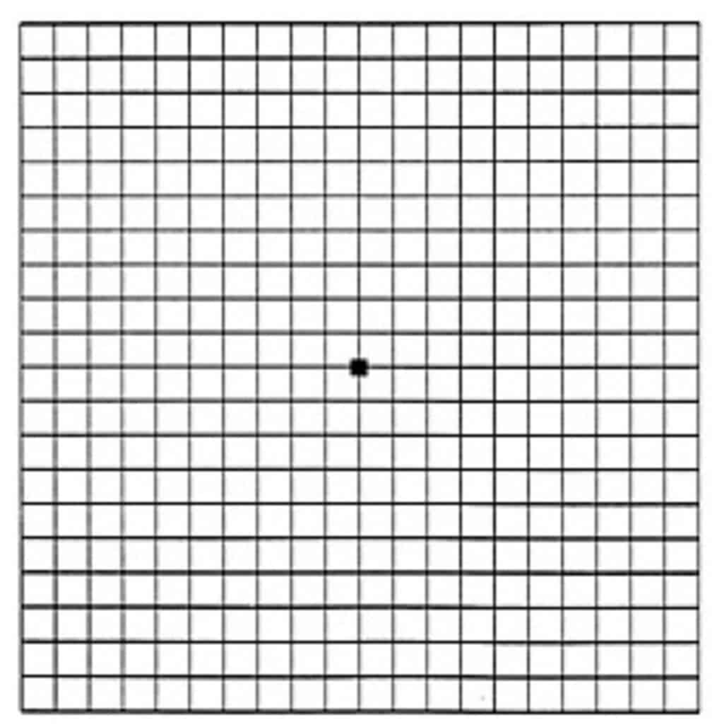
What is the Amsler Grid?

Those with age-related macular degeneration should be familiar with the Amsler Grid.  The grid is a cheap and easy method for self-monitoring vision changes in the central 20° of vision.   The grid is usually given by the examining eye doctor to those patients who have macular changes.  Traditionally, it is a piece of paper or card with the grid printed on it.  

A Few Tips for Observing the Amsler Grid

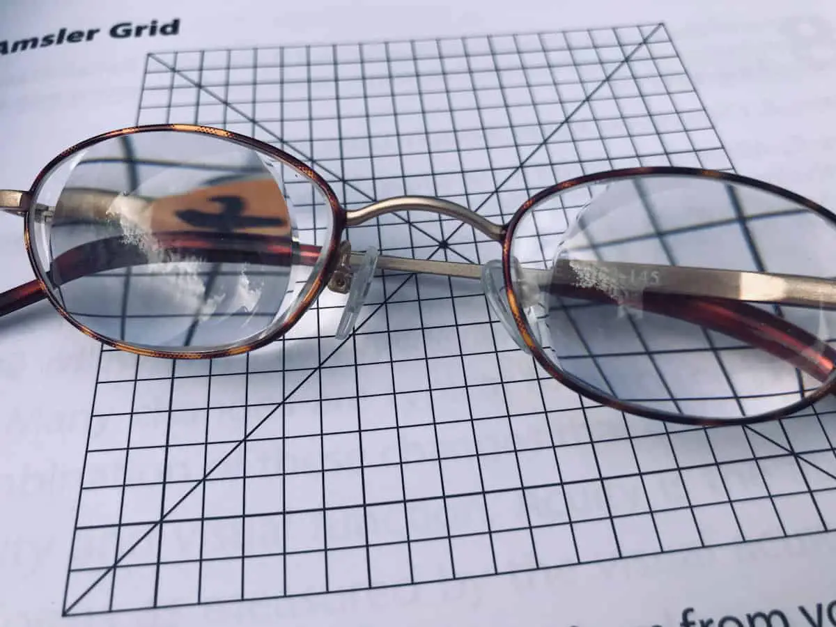
The grid is a 10 cm by 10 cm square, that is held 28 to 30 cm from your eye.   (This is about 12 inches.)  It is best to do the test in an area with good lighting.  You  should wear your best-correcting eyeglasses for near reading. 

Test one eye at a time.  A change in the eye to be tested will not be noticed if compensated for by the other open eye. 

This is an observational test of the macula and the visual area surrounding it.   With one eye covered, the open eye focuses on the center dot.  Resist the urge to look around the grid. 

Observe: while looking at the center dot;

  • Can you see the four corners and four sides?
  • Are there any areas where the lines and squares are missing, doubled, or blurry?
  • Are all the lines and squares straight?  Any waviness or distortion?

It is recommended, if you note a change from the previous self-testing, to make a note or mark on the grid where the change is located and call your retina doctor.

What Do Changes to the Amsler Grid Mean?

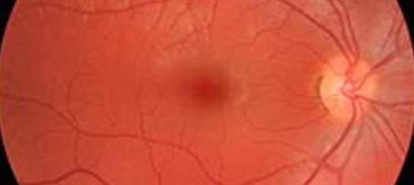
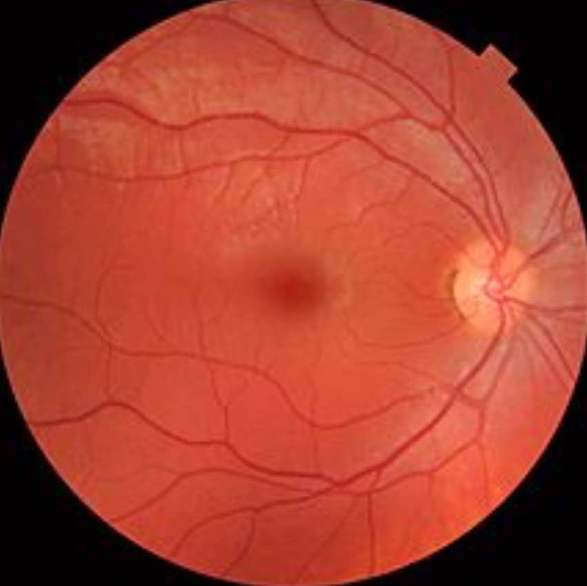
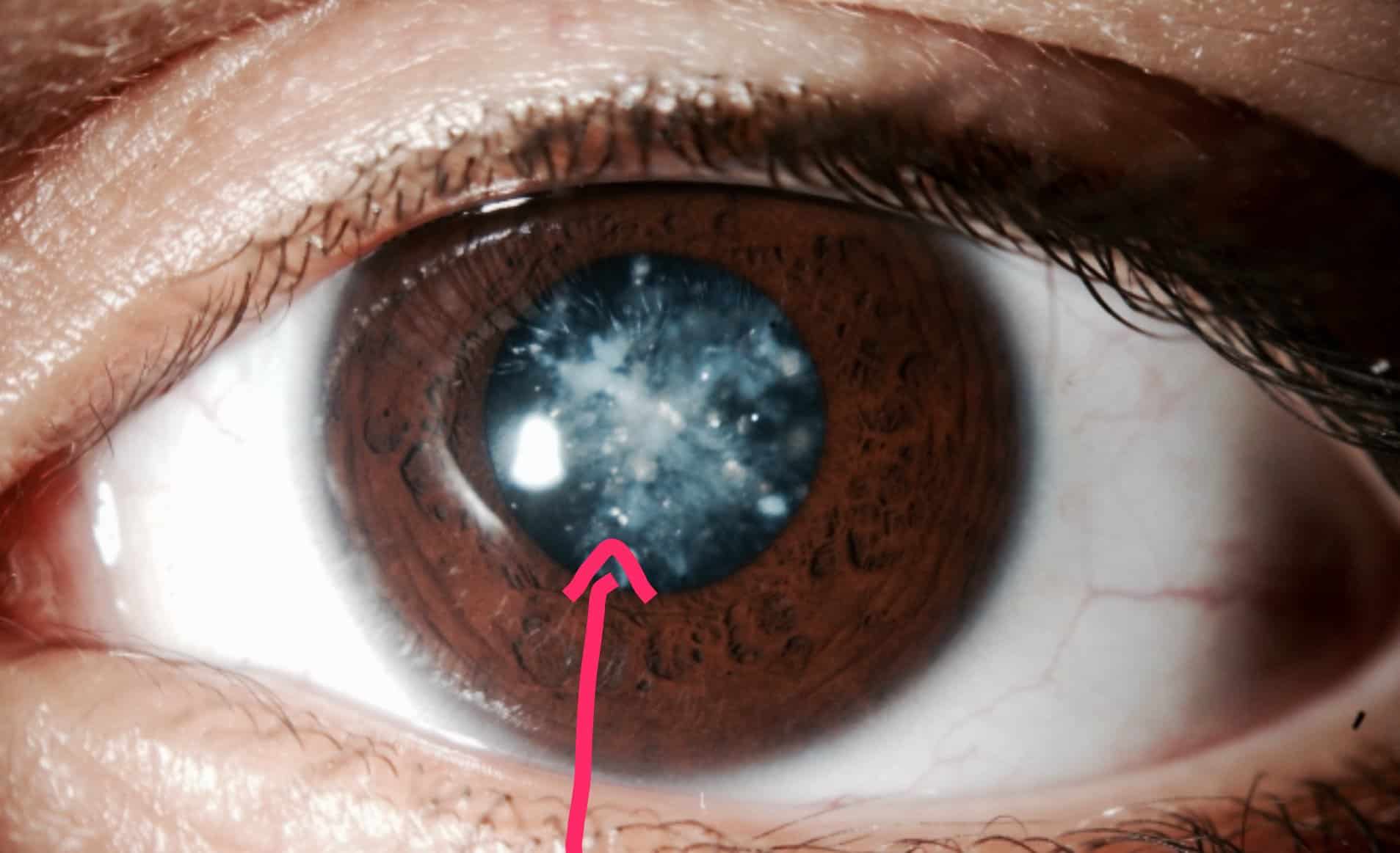
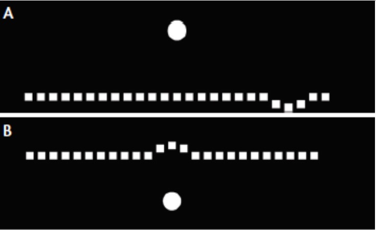
Metamorphopsia.  If you have dry macular degeneration or diabetic retinal disease, you may notice distortion of the central area (Image, above right.)  This indicates a change to the macula in which the photoreceptors (light-sensitive neurons) have become displaced.  This displacement could indicate the onset of the wet form of macular degeneration or diabetic macular edema. 

Other pathologies that cause distortion: cystic macular edema and epiretinal membranes.   This warrants additional testing by a specialist of retinal disease. Remember, early detection is key to the preservation of the central vision.

Missing or blurred areas.  An area that is missing or blurred can indicate the presence of a scotoma.  A scotoma is a blind area surrounded by normal areas and is caused by a degenerative disease.  This blind area can occur in the central macular area.   A scotoma of the central area will make it difficult to focus on the central dot of the grid.  A grid with guidelines can help to find the central area to align the eye.

If the scotoma is in an area like the corner or one of the sides, it could indicate visual field loss.  Visual field losses can indicate another disease process like retinitis pigmentosa, glaucoma, or pituitary tumor.

While the Amsler gird can be beneficial for detecting vision changes that  otherwise might not be noticed, it is not perfect.  It is not very sensitive.  Room lighting, the distance the chart is held, and adequate near vision with a set of optimal reading glasses can determine if the test is effective in detecting macular changes.  I have found the lack of patient compliance to also be a determining factor.  Can’t find it if you don’t look.

What are other methods of Self-Monitoring for Retinal Disease?

Is there an App for that?  Yes.

Amsler grid-based tests can be found in the  Apple (App Store) and Android (Google Play) app stores. Just type in ‘Amsler grid.’

There are two categories of self-monitoring apps.

1. Independent self-monitoring and

2. Apps with registration to a monitoring service.  The service in turn connects to an eye doctor’s office.

Independent monitoring, apps that do not connect to an eye care provider required.

I took a look at a few self-monitoring apps that I found in the Apple App store.  Google Play also has several apps.

AMD eye app store icon

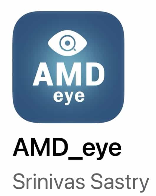
AMD eye  (for iPhone, iPad)  This app was designed by an ophthalmologist.  It is probably the most simplistic of the ones I tried.  The grid does not have the standard ‘square’ appearance. There does not seem to be a way to mark up or keep a record of what is observed for future comparison.  The user does have the capability to schedule daily or weekly alerts as reminders to test.  There is also some macular degeneration education data on the app. 

Amsler grid by Konan app store icon

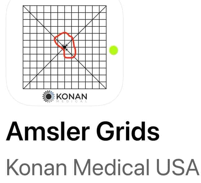
Amsler grids by Konan.  (for iPad) This app was developed by a designer of medical diagnostic instruments.  The app uses large print icons, has several types of grids to view, and has the ability to mark up the grid for future comparison. The marked-up grid can be labeled and saved to photos. Uniquely, it has a blind spot test to help the user establish a consistent testing distance.

The print on the instruction page is very small.  A low vision patient would benefit from initial instruction on how to use the app.  I would remind the developers that not everyone knows what OD and OS indicate. (OD = right eye, OS = left eye.)

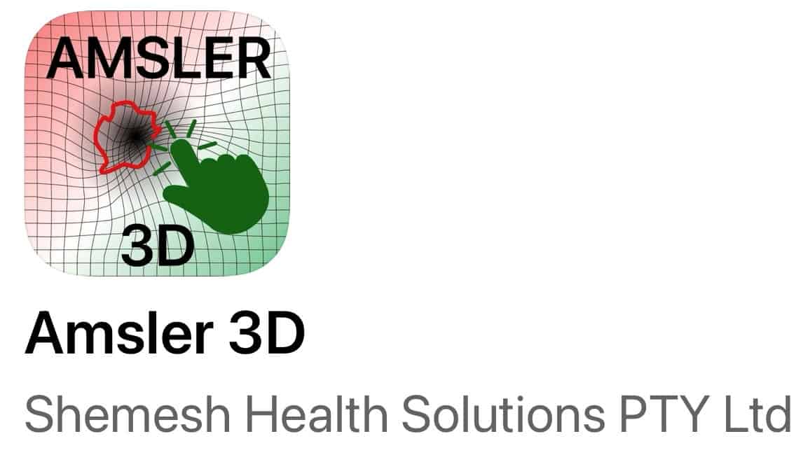
Amsler 3D. (for iPad)  Uniquely this app has an audio coach instructing the user on the correct testing procedure.  It has several grid-type options including a flashing center dot option.  The user has the ability to mark up the grid, label it with the date and save it under the setting indicated as Reports.  The report can be emailed to a doctor or saved as a photo.

LooC app store icon

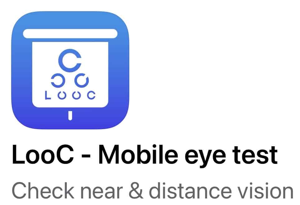
LooC (for iPhone, iPad)  This app uses an optotype called the Landolt C to test distance vision, near vision, color vision, and has an Amsler grid. It is interactive and uses an audio coach to give instructions. Results from the tests can be saved and shared.  

Maculatester app store icon

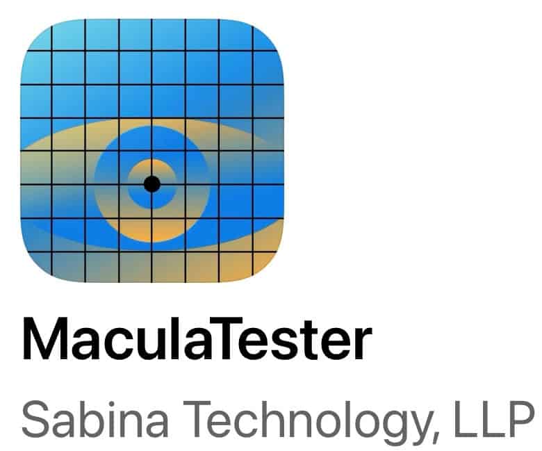
MaculaTester  (for iPhone, iPad, and Android) The grid for this app does not look like the standard Amsler grid, it is rectangular and fills the screen. It is very straight forward with not a lot of options.  The print is very small on the instruction screen and the testing screen.  The user can mark up the grid to save and share. (This is the only one I paid for.)

MAVA app store app

MAVA Mobile Acuity and Visual Assessment.  (for iPhone, iPad)  This is a very simple app, without options.  The user can mark up the grid, identify which eye, and save and share it.  That’s it.

The app I am more likely to recommend to my patients is the Amsler 3D.  I liked the audio coaching to remind the patient of the correct technique for testing and which large icon to tap next.

Apps connected to eye care professionals:

Alleye app store icon

Alleye (for iPhone, iPad, Android)This app requires a recommendation by an eye care physician to access the app.  The app should be installed at the eye doctor’s office, where it can be demonstrated.

The test does not use the familiar Amsler grid but does use an interactive task where the user moves dots into positions along a line.  The test data is encrypted, transmitted, and stored by Oculocare Medical.

Odysight app store icon

OydSight. This app uses gaming technology to engage users for self-monitoring between eye doctor visits.  The developer warns that it requires some cognitive ability. It utilizes visual acuity and an Amsler grid.

ForeseeHome Device for Self Monitoring

ForeseeHome tabletop monitor

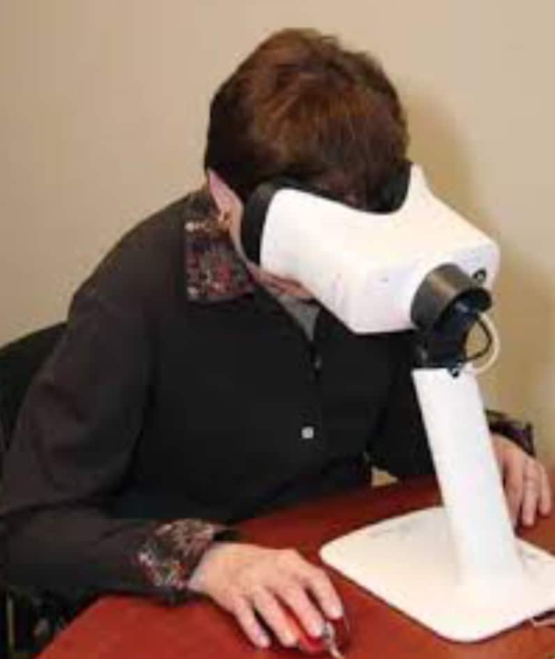
The device is called ForeseeHome by Notal Vision.   It is a table-top monitoring device to be used daily.  The test takes about 3 minutes per eye.  The test results are sent to Notal Vision.  If changes are detected, Notal Vision notifies your eye care doctor.  The plan is for  your doctor’s office to call to make an appointment for you to see the doctor.  In this way, you can receive prompt treatment to preserve your vision.

The Foresee Home overcomes some of the pitfalls of the self-monitoring Amsler grid.  This technology is more interactive.  The user knows that the results will be looked at and they become accountable for getting it done.  It is also more standardized because lighting, head position, and eyeglass prescription are not critical for test viewing. It is designed to detect the visual distortion (metamorphopsia )of wet macular degeneration.  It does require stable central vision and good cognitive function.

imaage of the ForeseeHome dot test
ForeseeHome test screen

How to get hooked up with the ForeseeHome system:

First, you must be identified by your doctor as being at risk for developing wet AMD.  It is also the first step to getting help through insurance to get coverage for the system.  Their website indicates that Medicare will cover some of the costs. 

Follow this link for doctors in your area who participate in this Notal  Vision program: Find a DoctorOpens in a new tab. 

The setup is quite easy.  You do not need a computer or even wifi.  The Notal company has a phone number to help you get set up. 

For more information on how it works, follow this link:  ForeseeHomeOpens in a new tab. 

Learn more about macular degeneration from my other article:  The 7 Truths about AMD

In the End…

Self-monitoring is a way for those at risk for progressive eye disease to conceivably pick up changes in vision that might otherwise go unnoticed.  It is a tool to be used in-between eye examinations and not a replacement for comprehensive eye care. 

The paper version of the Amsler grid has been used with limited success for over 100 years.  Its longevity indicates the importance of self-monitoring for the early detection of central retinal disease.  It is inexpensive and easy to do with little training.

The Amsler grid testing is not perfect.  The use of digital apps and home testing devices are more interactive and testing can be saved and shared.  Because of this, new technologies have the potential to engage users to be more compliant with regular self-monitoring tests.  The limitation is that many seniors may not have access to technology, may not understand its use, or may not have the physical or cognitive capacity to use smartphones and tablets. 

How Paleo, Vegetarian, and Gluten-free Diets Can Affect Eye Health


3  diets, Paleo, vegetarian, and gluten-free, which eliminate certain foods, how they may impact eye health.

Deprived of an adequate amount of basic nutritional building blocks results in defects of function, premature aging, disease, and decreased energy reserves. What ever effects the body, has an impact on the eyes. 

Eating can be enjoyable, least we forget that eating provides us with the nutritional building blocks of vitamins, amino acids, fats, and minerals needed to maintain a healthy f body.

The three main food groups are proteins, carbohydrates, and fats. Proteins are the building blocks of body tissue, things like muscle, collagen, and epithelium. These tissues are constantly turning over and renewing themselves in a healthy body. 

Carbohydrates fuel the body, not just for exercise, but also for maintaining cellular  function.  Excess carbohydrates are stored as fat. 

Dietary fats are the oils and solid fats found in animal and vegetable sources.  They are necessary for nerve development, as a source of stored energy, and help with vitamin absorption. 

Additionally, foods also supply us with vitamins and minerals:

Vitamins are organic micronutrients, made by plants and animals, that are essential to many cell functions in the body.  They serve as anti-oxidants,   help with cell growth and development,  integral in cellular metabolism,  and energy transfer.  Many of these vitamins can not be produced by our bodies and must be ingested from food.  

Deficiency of these micronutrients results in skin changes, deficits in neurological function, metabolic disorders,  decreased energy, and changes in vision.

Minerals are the inorganic molecules found in our foods.  They are also important for energy transport, bone health, nerve function, oxygen transport, and cellular metabolism, among other functions.   Minerals are integral to so many food s that a healthy diet should not exclude any one mineral. 

3 Healthy Diets

The trendy, healthy diets tend to manipulate the proportions or types of the 3 food groups.  The concern is not only the reduction or elimination of one or more of these groups, but the vitamins and minerals that come along with the foods that we eat.

Each of the 3 diets listed here are in effect, healthy diets.  In no way am I suggesting to avoid a diet that can improve your health.  Just be aware that eliminating certain foods from your diet can leave a gap in nutrition. The suggestion is to find alternative sources and/or supplement.  Consult your doctor as to the best alternatives.

The Paleo Diet

You can call this the Caveman diet.  It eliminates processed foods, that means: no wheat, sugar, beans, dairy, or alcohol.  It is a diet high in proteins and fats, and lower in carbohydrates.  The only source of carbs should be fruits and vegetables.  The healthy aspect is high fiber, lots of anti-oxidants, and lower blood sugar levels.  A good choice for those who are diabetic or pre-diabetic.

How a Paleo Diet might effect the Eyes

Since there is no dairy consumption there may be a decrease in vitamin D levels.  Vitamin D helps to maintain blood vessel integrity.  The central vision area , called the macula,  receives oxygen and nutrients from a blood vessels that are small and delicate. Impaired vascular health and blood vessel inflammation can increases the risk for macular degeneration.  Those on the Paleo diet should consider supplementing with Vitamin D.

Vegetarian and Vegan Diets

The vegetarian/vegan diet s are actually a broad category of specialized diets. Vegetarians usually will not eat any animal flesh, but there are some who eat fish.  Within this category there are those who will not eat dairy and/or eggs. 

Vegans are the strictest category that eliminates any animal products.  The benefit of vegetarian diets is lower body mass and cholesterol levels, meaning lower rates of cardiovascular disease and diabetes. Both of these benefits are good for the eyes.

How a Vegetarian Diet may Effect the Eyes

Most vegetarians I have met are usually aware of the nutrition deficiencies of their dietary choice. There are many nutrients and vitamins found in animal products: protein, iron, zinc, calcium, vitamin D, riboflavin, vitamin B12, vitamin A, omega-3 and iodine.  Each one of these plays an important role in our general health, and therefore, our eye health. Deficiencies can result in night blindness, vascular insufficiency, dry eyes, inflammation, and nerve damage. 

Since this is the most restrictive of diets, a vegetarian must be informed and aware of their diet deficiencies.  They must be careful to maintain a balance of alternative food sources and add supplements to their diet.

Gluten-Free Diet

Taking on a gluten-free diet is not usually a choice.  Commonly individuals find that by eliminating gluten or wheat products from their diet, they enjoy feeling better. 

There are actually 3 categories of individuals who have a negative response to gluten or wheat proteins: non-celiac gluten sensitivity, wheat allergies, and Celiac disease.  The symptoms associated with all three of these forms of gluten sensitivity are similar: bloating, diarrhea, headache, fatigue, generalized joint and muscle pain, and ‘brain fog.’  Celiac disease differs because it is an autoimmune disease (meaning it is genetic, not acquired) that can result in damage to  the gastrointestinal tract.  A damaged GI tract results in poor absorption of nutrients and vitamins. These deficiencies can result in weight loss and other diseases, like osteoporosis, skin disorders, and neurological disorders.

How a Gluten-free Diet can Affect the Eyes

Different from the other healthy diets, eliminating gluten is the only treatment for the signs and symptoms of gluten sensitivity. Those who have Celiac Disease and do not eliminate gluten are in for more health and eye related problems than those with non-Celiac gluten sensitivity and wheat sensitivity.  The mal-absorption of nutrients and the deficiency of anti-oxidants can result in cataracts, dry eyes, retinopathy, and night blindness for those with Celiac disease.

As a side note to those with gluten sensitivity and wheat allergies, some  lotions and makeup products have wheat in them, which can result in eye irritation and eczema when applied to the skin and eye area.

-Learn ore about supplementing for eye health in my other article: What is the Best Supplement to Preserve Eye and Brain Health?

In the end…

There are many reasons to change dietary habits. Commonly it is for weight loss, but for those with diabetes, heart disease, or Celiac disease, it maybe a necessity. Be aware of nutrient deficits in your diet. Certainly it is best to get our vitamins and minerals from good food sources rather than a pill.

If you have evaluated your diet and feel you need to supplement, choose wisely. There is such a thing as over- supplementing with nutrients that you don’t need. Also be wary of taking too large a dose of an essential nutrient. Read the supplement bottles carefully. If you have concerns get advice from your doctor or a registered dietician.

Meso-Zeaxanthin: The Third Carotenoid for Macular Health


Studies have shown that it is not just lutein and zeaxanthin that provide support to the retina, but there is a third carotenoid, meso-zeaxanthin that needs to be present for optimum anti-oxidant effect.

Lutein, zeaxanthin, and meso-zeaxanthin  are characterized as he yellow pigment concentrated in the macula of the retina and collectively are called macular pigment.  They  provide protection, by the absorption of blue light and anti-oxidant activity, and thereby aid in the protection and function of the macula.

Studies have shown that there is a drop in macular pigment decades before the onset of  macular degeneration.  Those studies where participants took lutein and zeaxanthin experienced an increase in the vital macular pigment.  Those who also supplemented with meso-zeaxanthin saw an even greater increase in macular pigment, more so than those who did hot supplement with meso-zeaxanthin.  (Ref: Lutein, Zeaxanthin and Meso-zeaxanthin Supplementation….)

There is a bonus to supplementing with the carotenoids; multiple studies have found that test subjects who supplemented with carotenoids not only increased macular pigment, but also experienced improved cognitive function.  Even young college students in one study benefited from carotenoid supplements. The trend is that eye doctors are now recommending to their patients who have macular degeneration and those at risk for developing MD, supplements which include lutein, zeaxanthin, and meso-zeaxanthin.

The Age-Related Eye Disease Study 2 (AREDS2) was a 5 year study  published in 2013.  Notably, in this second study, was the addition of two carotenoids as part of the supplement group.  The carotenoids are lutein and zeaxanthin.  The conclusion of the study was that these two carotenoids helped limit the progression of AMD.

Scientists indicate that meso-zeaxanthin is made from lutein.  The question arises; Have you ingested enough lutein and is it converted to the  meso-zeaxanthin form in sufficient amounts to make enough macular pigment to provide protection to the macula?

Food Sources of Meso-zeaxanthin

Although it is thought that some of the meso-zeaxanthin is derived from lutein, the rest needs to be derived from foods. Food sources are mainly, whole grains, egg yolks, and fatty fish (Rainbow trout and salmon, especially the skin).  We can never be really sure how much of the vital carotenoids we are getting, even from what we think is a nutritious diet.

In an age where our foods are produced by farm “factories” and may be genetically modified, the nutritional value may not be what we expect.  That is where supplementation becomes important for those with critical needs to maintain ocular and general health.

What is the difference between zeaxanthin and meso-zeaxanthin?

Meso-zeaxanthin is a geometrical diastereomer of zeaxanthin. (Definition of diastereomers: are molecules that are mirror images of each other.)  Because they are isomers they will have different biological  and functional prosperities for light filtering and anti-oxidant  capabilities.   (Ref: What is meso-zeaxanthin, and where does it come from?  )

Meso-zeaxanthin Supplements

Supplementing the diet directly with MZ (safely) can prevent the macula from becoming deficient.  The meso-zeaxanthin, found in supplements, is formed from natural lutein.  It is extracted from the Aztec Marigold flower.  These are the extracts which are most frequently used in clinical trials.

orange and yellow marigolds

A note for those taking beta-carotene supplements: Biochemistry is complicated.  The AREDS 2 study found that the beta-carotene had the capability of competing with, and therefore decreasing, the absorption of lutein, zeaxanthin, and meso-zeaxanthin.  So do not take them together.  (Note; AREDS supplement formula has beta-carotene, whereas AREDS2 does not have beta-carotene, but does have lutein and zeaxanthin).

Safety of Meso-zeaxanthin

Several studies showed no observed adverse side effects in dosages of 200 or 300 mg/kg body weight.  This dosage is far beyond dietary and commercially available supplement dosages which are usually 2 – 4 mg.

Be aware, when shopping for eye vitamins, the most popular AREDS and AREDS2 formulas do not include meso-zeaxanthin.

Learn more about the carotenoids and their importance to eye health, in my other article: Carotenoids Needed for Eye Health, the Essentials

In the End…

While macular pigment has been determined to be comprised principally  of the three carotenoids; lutein, zeaxanthin, and meso-zeaxanthin, very little is known or understood regarding the role of meso-zeaxanthin. Studies have indicated that its inclusion in macular pigment may be essential for optimal macular protection and function.

Preventing Vision Loss from Age-Related Eye Disease


photo of the retina of a healthy eye

Studies have shown that intervention with lifestyle changes which includes: adequate nutrition with supplementation, exercise and weight loss, and reducing environmental stresses like UV exposure and smoking can halt the progression and prevent further vision loss from age-related eye diseases.

How do you keep your eyesight from deteriorating with age?

There are 4 major risk factors for developing eye disease which can result in vision loss:

  • a nutritionally poor diet,
  • lack of exercise,
  • smoking, and
  • UV and high energy blue light exposure.

We are living longer than our ancestors did less than one hundred years ago.  Diseases associated with old age in the past were rare, like cognitive impairments, cardiovascular disease, and some types of cancer, but are now more common in an aging population.  It can also be said that macular degeneration is on the rise because of the increase number of those living to be older, the Western diet, and environmental stresses.

What can be done to decrease the risk for age-related eye diseases?

Normal retina: central macula and optic nerve

Food be thy medicine and medicine be thy food.” Hippocrates

Healthy body, healthy eyes.  The greatest problem we have as a western society is our lack of fresh and unprocessed foods.  Life is easier with foods processed and prepared by commercial food producers.  The problem is that nutrition is often processed out of the food, or replaced by tasty, fatty, and are high carbohydrates that are low in nutritional value.

The end result is obesity accompanied with diabetes, cardiovascular disease, and metabolic syndrome. All systems of the body are susceptible to the degenerative  effects of poor nutrition, including the eyes.  It is these systemic conditions that contribute to  cataract formation, macular degeneration,  diabetic retinopathy, and glaucoma.

Macular degeneration with drusen

Ideally, our doctors would like to know that their patients are eating healthy and nutritious foods for both ocular and general medical health.  What the famous AREDS (age-related eye disease study) studies showed us is that nutrition makes a difference.  In the case of this study, the improvements were modest but improvements nonetheless.  The importance of this study, and others like it, indicate that specific nutrients do make a difference in our overall health. 

Eating healthful foods, with adequate

Diabetic retinopathy with hemorrhages

nutritional proportions, is not always easy It is the rare individual who spends time evaluating the quality and nutritional benefits of the foods they eat each day.  What we eat is limited by time of year, geography, preferences, and availability.  That is where supplements come in.  Not everyone eats fish (for omega 3s) or likes leafy green vegetables.  What you need to do is to look at what may be missing in your diet.  Talk to a dietitian or a naturopath.

The AREDS 2 study supplement included:

  • Vitamin C
  • Vitamin E
  • zinc
  • copper
  • lutein
  • zeaxanthin     

Recent and ongoing research has identified other nutrients beneficial to ocular health.  

  • other carotenoids, like Astaxanthin,
  • flavonoids, like the berry anthocyanin extracts,
  • resveratrol which is a polyphenol, and
  • saffron extract.

See these other articles for more details on the impact other nutrients can have on ocular health:

How do AREDS and AREDS2 Differ?

Do Omega-3 Supplements Help Your Eyes?

Astaxanthin and Vision Benefits

Eye Vitamins; The Anthocyanins of Bilberry, Blueberry, Maqui berry, and Black currant

Resveratrol

Saffron and Eye Health

How adequate nutrition with supplementation serves us 3 ways:

  1. Optimize your vision and physical health,
  2. disease prevention, and
  3. reduce progression of macular disease.

Lifestyle Changes

The concept of movement as exercise is nothing new.  Many athletic types have made lots of money showing us how to exercise.  TV doctors and talk show hosts all expound on the benefits of exercise.  It does make a difference both for eye health and general health.  Many health problems such as  high blood pressure, diabetes, and high cholesterol are linked to eye disease.  What is good for the body is good for the eyes.

How Exercise Effects Eye Health

Exercise increases blood flow, resulting in more blood perfusion to the eye.  This can help reduce the risk for glaucoma and increase oxygen and nutrients to a macula which may be degenerating.  Those suffering from diabetes are encouraged to exercise to control blood sugar, which in turn helps to reduce the risk for diabetic retinopathy.

Stop Smoking!

Here we go…another reason to stop smoking.  Quitting smoking reduces your risk for some eye diseases.  This is the easiest risk factor to control. Those who smoke are more likely to develop cataracts at an earlier age.  Studies (Clinical Sciences) have shown that those who quit smoking decrease  the risk of developing cataracts that will require surgical removal.

The inhaled cigarette smoke is known to contain numerous toxic  compounds, some are known to cause cancer. These toxins cause oxidative damage, blood vessel changes and inflammation.(see Journal of Ophthalmology) It is known that smoking effects blood vessel function, and anything that effects blood flow will impact the very tiny blood vessels that feed the very sensitive macula.  Smoking increases the risk of macular degeneration.

UV Protection 

Cortical cataracts

We apply sun screen lotions with SPF (sun protection factors) to prevent sunburns to the skin.  Think of sunglasses as SPF for the eyes.  Good quality sun-glass lenses will protect against 100% of the tissue damaging ultra violet rays.  Bigger lenses are better, not only to protect the lens and retina of the eye, but also protects the delicate tissues around the eyes. 

How UV Light and High Energy Blue Light Affects Eyes

Studies have shown that chronic, long-term  UV light exposure can cause early

Non -cancerous conjunctival growth: pinguecula

onset of cataracts and macular degeneration later in life.  Exposure of the conjunctiva (whites of the eyes)and eyelids can result in growths and worst yet…cancers.

Visible light spectrum

The Blue Light Hazard

Both young and old who use technology chronically for long periods of time, as many of us do, need to be aware of the potential for eye damage due to exposure to blue light.  This is not the ultra violet invisible ‘light’, it is the high energy visible blue light.  Eye doctors are concerned that excessive exposure over an extensive period of time to high energy blue light will cause retinal degeneration.

Consider reducing time spent on digital devices.  For those who need to , there are computer screen filters which can be purchased to reduce blue light.  Eye glass lenses are made, which filter out specific wavelengths of blue light, thought to be the most damaging.  You would need to ask your eye doctor or optician for these specialty lenses.

Finally, there is an app for that!  Type in to your browser or app store:. blue light filter for…Apple, Windows, Android, Mac, Chrome, etc.  As a bonus, blue light filters can also help with computer eyestrain and difficulty getting to sleep after an extensive period of computer viewing in the evening.

It is not known yet how the eyes of the younger generation will fair after decades of chronic blue light exposure emitted from technology.  Medical professionals question their risk for retinal damage and macular degeneration.

In the end…

Both eyes and body benefit from a nutritionally healthful diet, exercise, and protection from environmental assault.  Scientific studies have proven that nutrition and lifestyle factors can promote health and decreases the risk for developing age related eye diseases that have the potential for vision loss and decreasing the quality of life for the aging population. 

You only have one set of eyes, take care of them.


The Solar Eclipse and Your Vision


NASA

Just a few notes about watching a solar eclipse. A patient called me a few days ago looking for solar eclipse glasses.  In my mind it is never a good idea to look in the direction of the sun.  I do realize, however, that there are a lot of people who want the experience of seeing a solar eclipse since it is so rare.

There is eye pathology associated with sun gazing called solar retinopathy. This damage is caused by high intensity light exposure.   Retinopathy is damage to the very sensitive  light detecting neurons in the back of the eye, which are responsible for vision.  While solar retinopathy will not result in total vision loss, it is damage to the macula, which is the area of our most critical and detained vision.

Looking at the sun, if inadequately protected, can result in solar retinal damage in 30 seconds or less.  Sunlight is not only a source of high intensity UVA and UVB wavelengths, but also radiates other potentially damaging wavelengths of light, such as Infrared and near infrared.  For this reason, regular  UV 400 protective sunglasses, no matter how dark, are not enough for solar eclipse watching.

Symptoms of solar retinopathy are felt a few minutes to a few hours after exposure, depending on the intensity of the burn. Visually there may be blurring, a scotoma (an area of no vision, a ‘blind spot’), and/or distortion accompanied by a headache.

The solar eclipse glasses which are recommended have additional specialty filters to guard against the other high intensity wavelengths of light. The international safety standard is labeled  ISO 12312-2.  This should be indicated on the glasses, along with the name of the manufacturer.  A list of reputable manufacturers of solar eclipse glasses can be found at American Astronomical Society(AAS), Vendors of Solar Filters and Viewers.  They are not expensive, but don’t take a chance on poorer quality, or even worse; fake solar filters.

If you would like a comprehensive description of eclipse viewing, go to the NASA.gov website.  On this page, they describe how to use the solar filters and make other safety recommendations. They also describe an indirect technique  to experience the solar eclipse.  Through the NASA.gov website you can access ways to watch live streams:

Solar eclipse, 2024: NASA Science .gov

Vision Loss, Hallucinations, and Charles Bonnet Syndrome


 Charles Bonnet Syndrome (CBS) is often regarded as a fascinating yet misunderstood occurrence associated with vision impairment. While commonly linked to those with vision loss, CBS encompasses much more than previously assumed visual hallucinations.                    

While Charles Bonnet Syndrome (CBS) is more commonly associated with elderly individuals, there have been documented cases of children experiencing visual hallucinations related to CBS.

However, one of the challenges with identifying CBS is that many individuals hesitate to report their visual experiences due to fears of being labeled as old, losing mental acuity, or being deemed in need of psychological intervention or medication.                            

Vladmir Kush art

Those who experience CBS hallucinations are those who:

  • have recent vision loss, not usually those with long term vision loss,
  • have lost the vision in both eyes, (20/100 or worse), and
  • are mentally alert, attentive, and understand the hallucinations are not real.

 Charles Bonnet Syndrome (CBS) predominantly affects the elderly, particularly those grappling with age-related eye conditions like macular degeneration. However, CBS isn’t exclusive to this demographic.

Individuals with vision impairments stemming from conditions like cataracts, glaucoma, diabetic retinopathy, optic neuropathy, or retinitis pigmentosa are also susceptible to experiencing CBS. Essentially, any condition causing vision loss along the brain’s visual pathway can trigger CBS episodes. 

Spectrum of Hallucinations in Charles Bonnet Syndrome

The hallucinations characteristic of Charles Bonnet Syndrome (CBS) manifest in various forms, ranging from simplistic lines and patterns to intricate scenes teeming with animals, people, and action. These visual experiences can take on a cartoonish appearance, be small and unobtrusive, or even exhibit grotesque features. Remarkably, they seamlessly integrate into the surrounding environment, appearing vivid and lifelike.

Duration-wise, CBS hallucinations vary widely, spanning from fleeting moments to prolonged episodes lasting hours. These episodes are episodic in nature, potentially persisting for months to a year before gradually subsiding.

While unsettling for those experiencing them, these hallucinations are typically not described as terrifying or menacing. Importantly, the visual images in CBS remain passive observers, devoid of interaction with the individual.

Research suggests that individuals living alone are more prone to experiencing CBS hallucinations. Furthermore, these episodes may be triggered by periods of quietude, low light levels, or exacerbated by fatigue and stress. 

Children and Charles Bonnet Syndrome

There have been documented cases of CBS occurring in children as well. While less common, children with various forms of vision impairment, such as retinal dystrophies or congenital blindness, have reported experiencing CBS hallucinations.

One study published in the British Journal of Ophthalmology examined CBS in children with visual impairment. The researchers found that children as young as five years old could experience CBS hallucinations, which ranged from simple patterns to complex scenes involving people and animals.

The study highlighted the importance of raising awareness among healthcare professionals and caregivers about CBS in pediatric populations.

Additionally, case reports and anecdotal evidence also contribute to our understanding of CBS in children. These reports often emphasize the challenges of diagnosing CBS in pediatric patients due to the difficulty children may have in articulating their visual experiences or the tendency for caregivers to dismiss hallucinations as mere imagination.

The Mechanisms Behind Charles Bonnet Syndrome Hallucinations

The visual hallucinations characteristic of Charles Bonnet Syndrome (CBS) are theorized to stem from sudden vision loss resulting from pathology within the visual system. This system encompasses the eye, neural connections, and the occipital cortex located at the rear of the brain.

Analogous to phantom pain experienced by amputees, these hallucinations emerge as the brain attempts to fill in the perceptual gaps left by the absence of visual stimuli. Just as an amputee may feel pain in a missing limb, individuals with CBS experience “phantom vision” where visual sensations persist despite the absence of external stimuli.

While the eye itself doesn’t register pain with vision loss, the nerves within the visual system continue to fire, generating images in the absence of genuine sensory input.

In essence, these hallucinations can be likened to phantom vision, offering a glimpse into the intricate workings of the brain’s perceptual processes amidst visual impairment.

Managing Charles Bonnet Syndrome: Coping Strategies and Support

In response to distressing hallucinations, individuals affected by Charles Bonnet Syndrome (CBS) may seek medical assistance. However, addressing these visual disturbances doesn’t entail a one-size-fits-all solution.

While resolving the underlying cause of vision loss, such as through cataract surgery for cataract-related vision impairment, can alleviate symptoms to some extent, restoring full vision is often unattainable as in macular degeneration, optic  neuropathy, or retinitis pigmentosa. 

Reports suggest that individuals have adopted various coping mechanisms to mitigate CBS hallucinations. These include techniques like eye movement, temporary eye closure, or adjustments in lighting conditions to dispel the hallucinatory episodes.

Additionally, efforts to reduce social isolation, engage in vision rehabilitation programs, and utilize optical aids play an important role in redirecting visual stimulation and improving overall well-being.

In the End…

Charles Bonnet Syndrome is a complex interplay between visual impairment and perceptual phenomena. Understanding the diverse manifestations of CBS hallucinations and the underlying mechanisms and coping strategies, it’s evident that CBS presents unique challenges for individuals grappling with vision loss.

By fostering greater awareness and support, we can enhance the quality of life for those affected by CBS and pave the way for more effective management strategies in the future.

Want to learn more about macular degeneration, read my other article: The 7 Truths about Macular Degeneration